Some of these figures and pages may be copyright protected. I take no responsibility for matters related to copyright laws and regulations. Use these at your own discretion.
Augustine DiGiovanna, Ph.D.
Videos
“Bone Matrix: Calcium Phosphate Salts and
Bone Growth”
https://blausen.com/en/video/bone-matrix-calcium-phosphate-salts-and-bone-growth/
“Bone Cells: Osteoclast Resorbs Bone and
Osteoblasts Form New Bone”
https://blausen.com/en/video/bone-cells-osteoclast-resorbs-bone-and-osteoblasts-form-new-bone/
“Compact Bone vs. Spongy Bone”
https://blausen.com/en/video/compact-bone-vs-spongy-bone/
“Synovial Membrane”
https://blausen.com/en/video/synovial-membrane/
“Osteoarthritis”
https://blausen.com/en/video/osteoarthritis/
“Osteoarthritis of the Hip”
https://blausen.com/en/video/osteoarthritis-of-the-hip/
‘Rheumatoid Arthritis – Knee”
https://blausen.com/en/video/rheumatoid-arthritis-knee/
“Rheumatoid Arthritis – Hand”
https://blausen.com/en/video/rheumatoid-arthritis-hand/
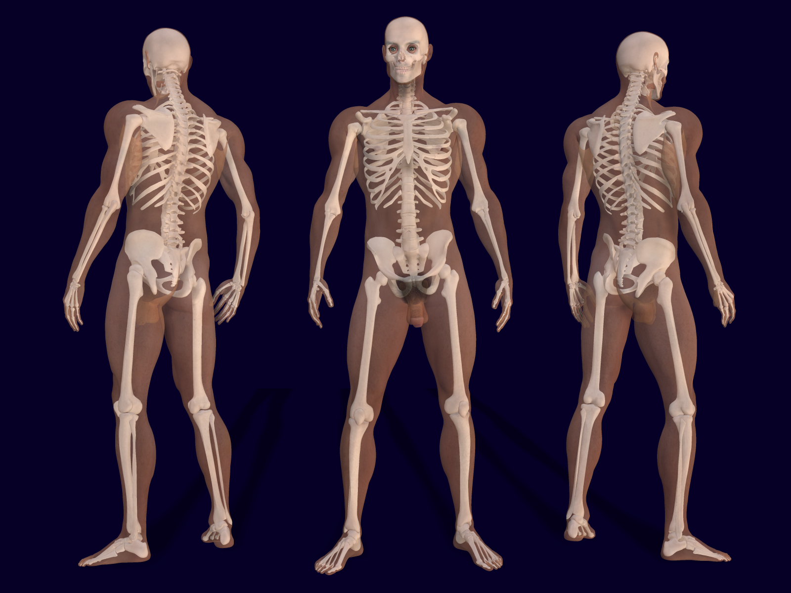
Fig.
9.1 The skeletal system.
https://upload.wikimedia.org/wikipedia/commons/b/b1/701_Axial_Skeleton-01.jpg
https://upload.wikimedia.org/wikipedia/commons/7/78/3D_Male_Skeleton_Anatomy.png
https://upload.wikimedia.org/wikipedia/commons/7/75/Human_skeleton_front_arrows_no_labels.svg
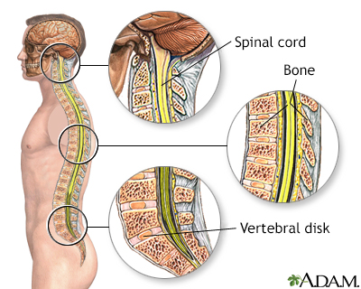
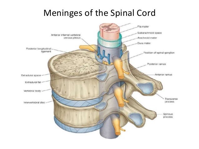
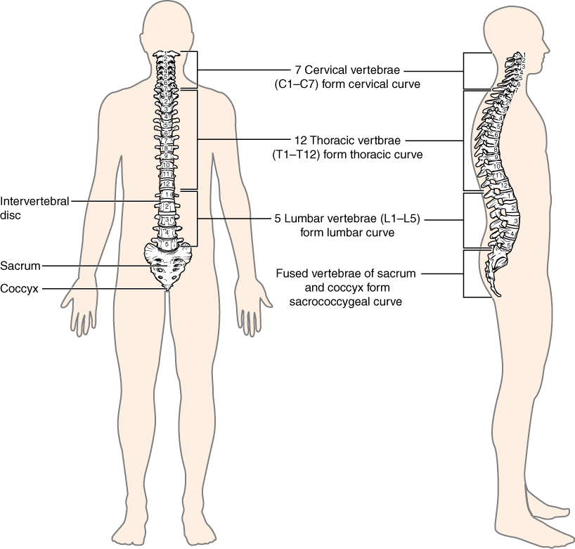
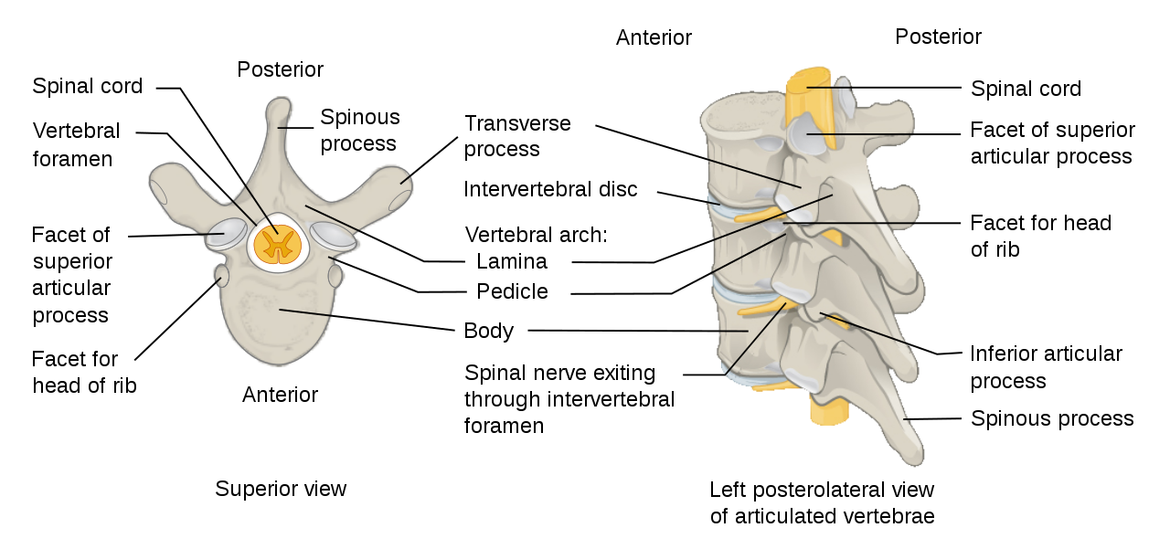
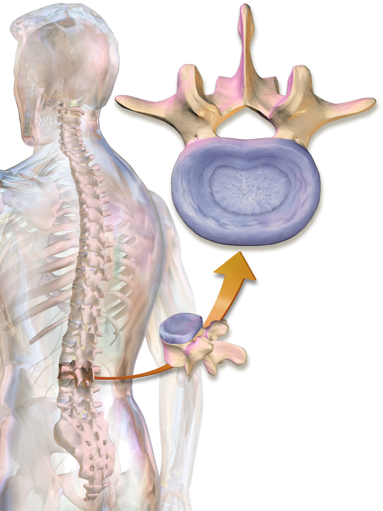
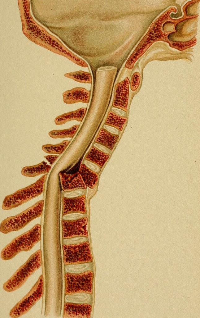
Fig.
9.2 Skeletal support for the spinal cord.
https://www.fairview.org/hlimg/krames/276757.jpg
https://ssl.adam.com/graphics/images/en/9561.jpg
https://upload.wikimedia.org/wikipedia/commons/8/82/715_Vertebral_Column.jpg
https://upload.wikimedia.org/wikipedia/commons/c/cf/Cervical_vertebra_blank.svg
https://commons.wikimedia.org/wiki/File:Cervical_vertebra_blank.svg
Fig.
9.3 Skeletal components as anchors and levers.
https://upload.wikimedia.org/wikipedia/commons/9/98/Anatomy_and_physiology_of_animals_Antagonistic_muscles%2C_flexion%26tension.jpg
https://upload.wikimedia.org/wikipedia/commons/6/6c/Musclesbicepstriceps_esp.jpg
https://upload.wikimedia.org/wikipedia/commons/1/1b/Relax1contract.jpg
https://commons.wikimedia.org/wiki/File:Relax1contract.jpg
https://upload.wikimedia.org/wikipedia/commons/2/23/603_Anatomy_of_Long_Bone.jpg
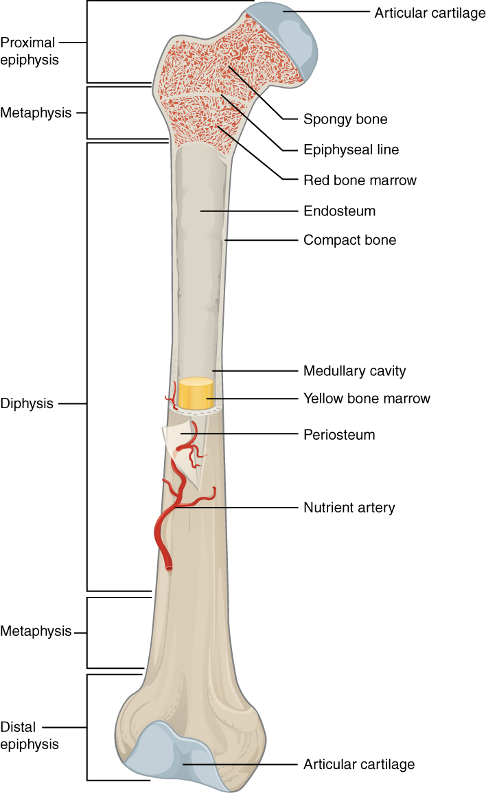
https://upload.wikimedia.org/wikipedia/commons/8/8f/Figure_38_02_04.png
Fig.
9.5 Bone tissue: compact bone and trabecular bone.
https://upload.wikimedia.org/wikipedia/commons/5/58/624_Diagram_of_Compact_Bone-new.jpg
https://upload.wikimedia.org/wikipedia/commons/4/49/Figure_33_02_09.jpg
https://upload.wikimedia.org/wikipedia/commons/f/ff/606_Spongy_Bone.jpg
Fig.
9.6 Age changes in bone tissue: (a) Trabecular bone. (b) Cortical bone.
https://commons.wikimedia.org/wiki/File:Osteoporosis_--_Smart-Servier.jpg
https://upload.wikimedia.org/wikipedia/commons/b/b8/Osteoporosis_6_--_Smart-Servier.png
https://upload.wikimedia.org/wikipedia/commons/0/07/Osteoporosis_02.png
https://upload.wikimedia.org/wikipedia/commons/a/af/Osteoporosis_Locations.png
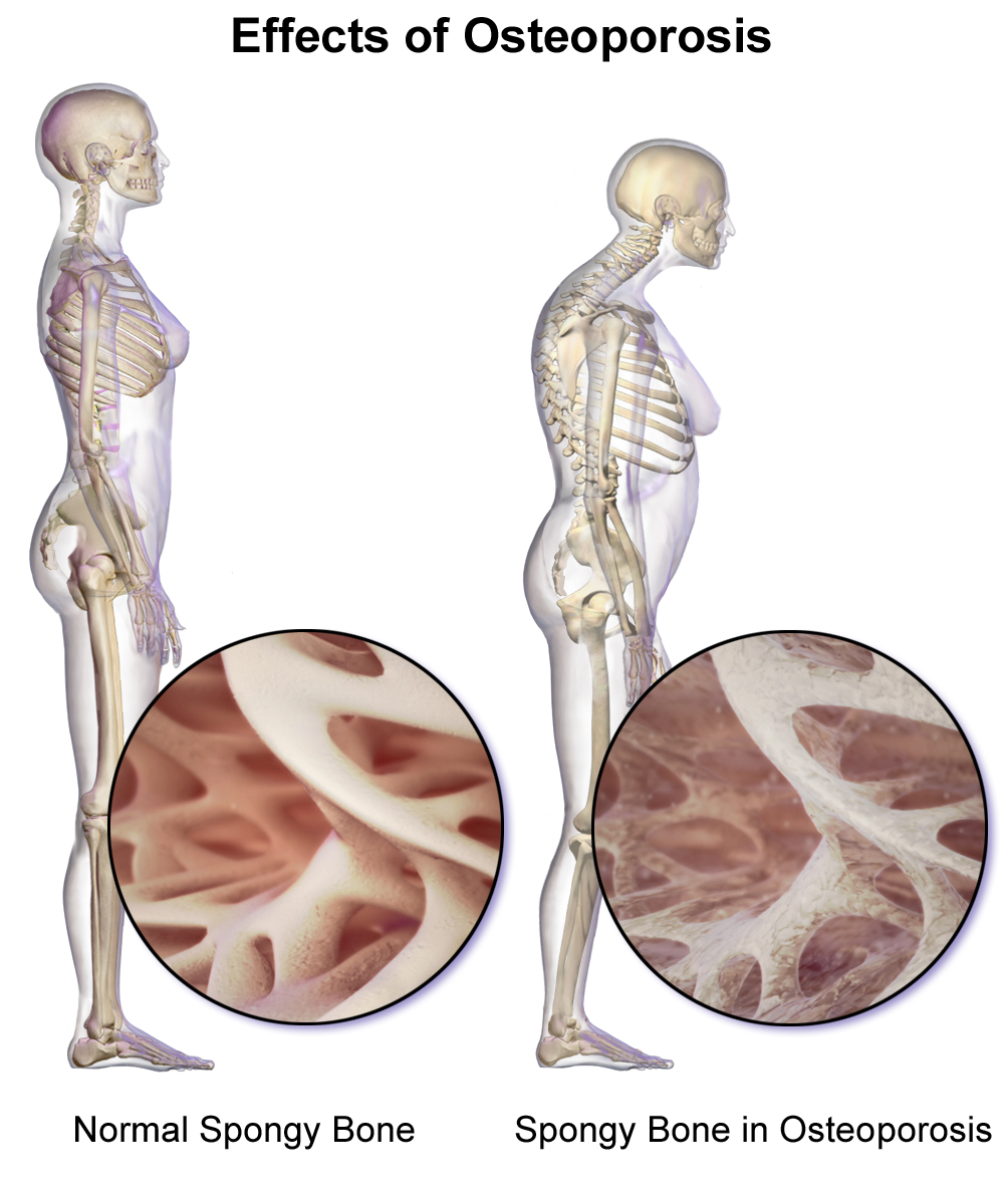
Fig.
9.7 Effects of osteoporosis on bone
https://upload.wikimedia.org/wikipedia/commons/1/11/Cdm_hip_fracture_343.jpg
https://upload.wikimedia.org/wikipedia/commons/2/2f/Skin_folds_over_a_hip_fracture.jpg
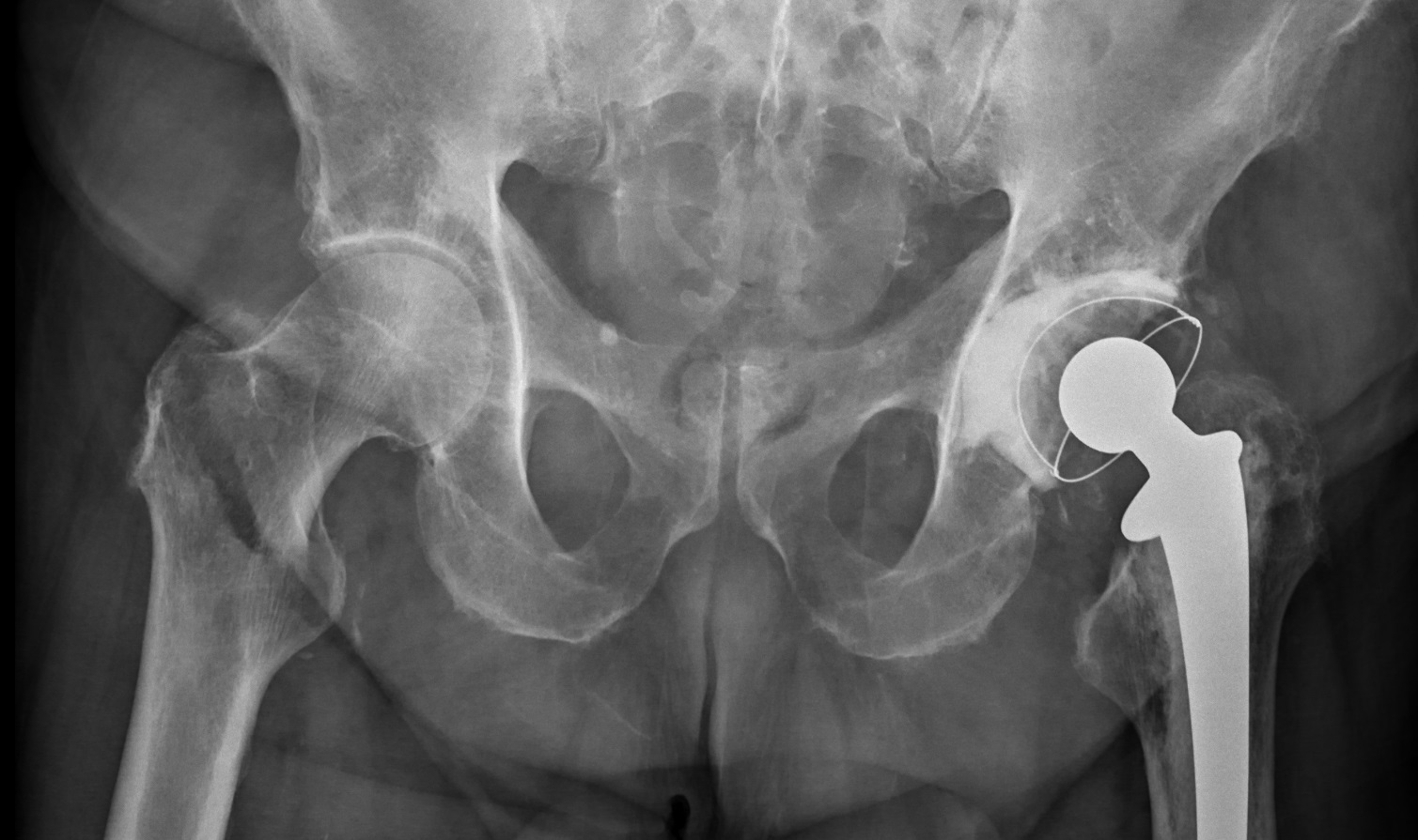
https://upload.wikimedia.org/wikipedia/commons/5/5c/Cdm_hip_implant_348.jpg
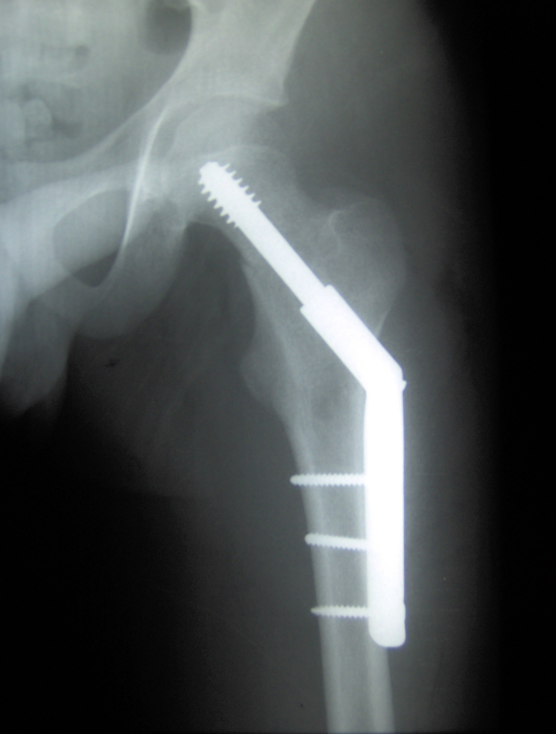
https://upload.wikimedia.org/wikipedia/commons/a/af/Osteoporosis_Locations.png
https://commons.wikimedia.org/wiki/File:Osteoporosis_--_Smart-Servier.jpg
https://commons.wikimedia.org/wiki/File:Menopause_-_Osteoporosis_--_Smart-Servier.jpg
https://upload.wikimedia.org/wikipedia/commons/e/e1/X-ray_of_stress_fracture_of_the_hip.jpg
https://upload.wikimedia.org/wikipedia/commons/d/dc/Colles_fracture.JPG
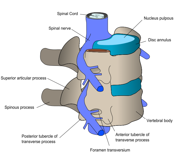
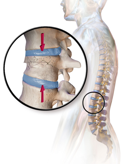
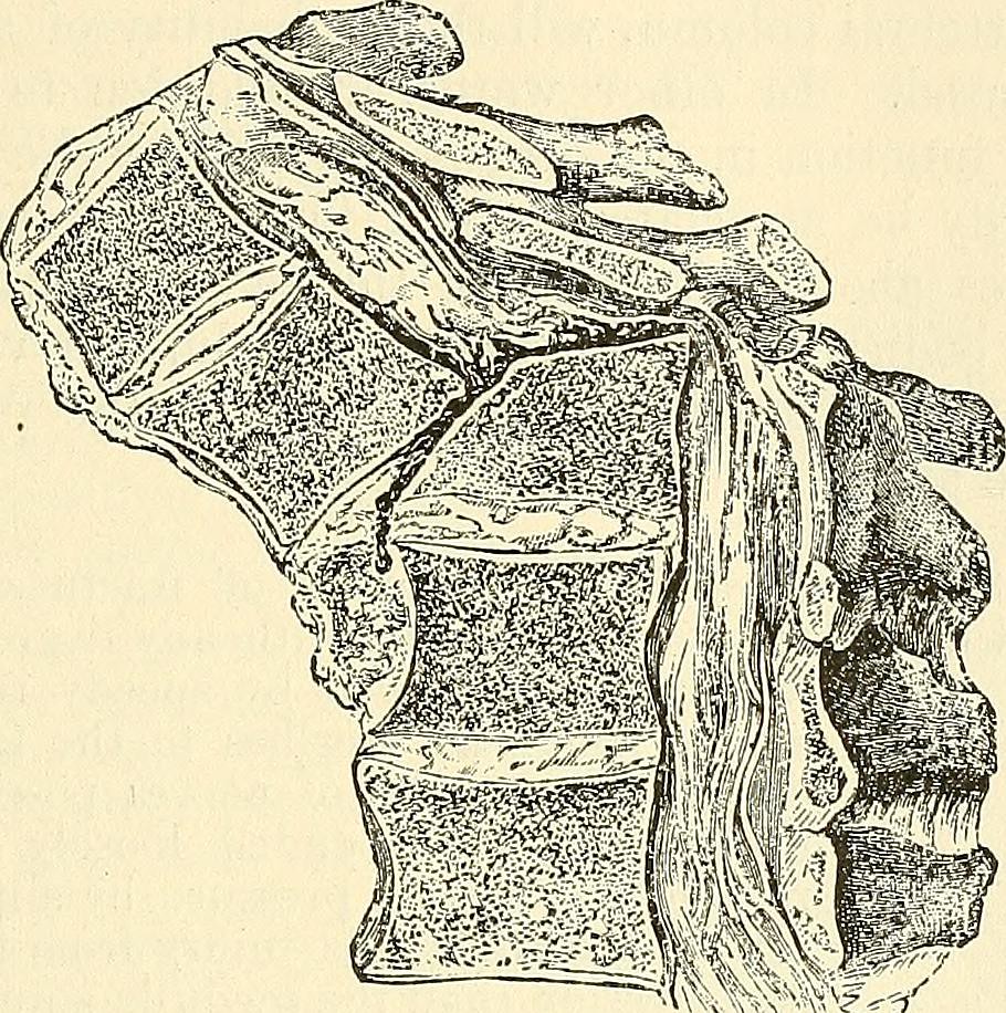
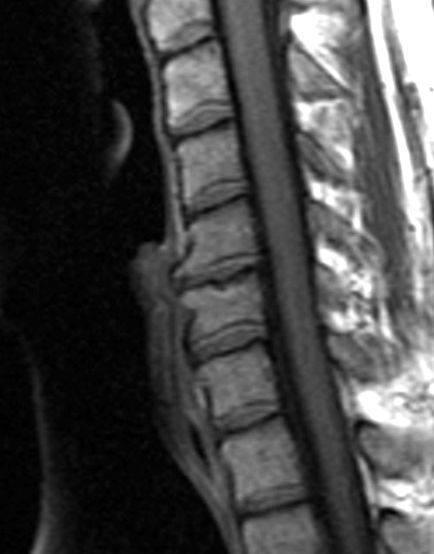
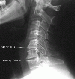
Fig.
9.8 Vertebrae and slightly movable joints: (a) Young vertebrae and joints. (b)
Crush fractures. (c) Age changes. (d) Intervertebral joints with
osteoarthritis.
(a)
Young vertebrae and joints.
https://upload.wikimedia.org/wikipedia/commons/1/1c/Annulus_Fibrosus.png
https://upload.wikimedia.org/wikipedia/commons/a/a4/Vertebra_Posterolateral.jpg
https://upload.wikimedia.org/wikipedia/commons/0/0f/716_Intervertebral_Disk.jpg
https://upload.wikimedia.org/wikipedia/commons/d/d9/Cervical_vertebra_english.png
https://live.staticflickr.com/306/20157149478_0eef48518d_b.jpg
https://live.staticflickr.com/2911/14784720293_5725cd4946_b.jpg
(b)
Crush fractures.
https://upload.wikimedia.org/wikipedia/commons/1/17/Blausen_0250_CompressionFracture_Vertebrae.png
https://live.staticflickr.com/3780/20336691242_30ea809a30_b.jpg
https://live.staticflickr.com/5570/14594772299_01806e31ac_b.jpg
https://live.staticflickr.com/5596/14593248120_eb911df60a_b.jpg
https://farm3.staticflickr.com/2917/14776795131_30672f6e98_b.jpg
https://upload.wikimedia.org/wikipedia/commons/c/c3/L1_2_vertebral_fracture.jpg
(c)
Age changes.
https://live.staticflickr.com/5573/14581563328_9845747bf7_z.jpg
https://live.staticflickr.com/3780/20336691242_30ea809a30_b.jpg
https://live.staticflickr.com/2903/14800430253_0326e6b47d_b.jpg
https://live.staticflickr.com/5559/14593907589_0421a0e421_b.jpg
https://live.staticflickr.com/2921/14593894989_a0259116f7_b.jpg
https://upload.wikimedia.org/wikipedia/commons/d/d3/728_Herniated_Disk.jpg
(d)
Intervertebral joints with osteoarthritis.
https://www.spinehealth.com/images/spinal-arthritis.jpg
VIDEOS
“Lumbar Osteoarthritis Video”
https://www.spine-health.com/video/lumbar-osteoarthritis-video
“Lumbar Spinal Stenosis : Diagnosis and Treatment Options”
See
the first 8 minutes
https://www.youtube.com/watch?v=YmeWKqhu-WM
https://www.youtube.com/watch?v=BR1xv5Gmvzw
https://neupsykey.com/spinal-cord/#cesec1
Fig.
9.8 Structure of freely movable joints.
https://ca.wikipedia.org/wiki/Articulaci%C3%B3_(anatomia)#/media/Fitxer:Joint_ca.svg
#/media/Fitxer:Joint_ca.svg)
https://upload.wikimedia.org/wikipedia/commons/thumb/e/e7/Joint_ca.svg/2000px-Joint_ca.svg.png
https://upload.wikimedia.org/wikipedia/commons/e/ed/909_Types_of_Synovial_Joints.jpg
Fig.
9.10 Osteoarthritis
https://upload.wikimedia.org/wikipedia/commons/4/42/X-ray_of_a_normal_hip.jpg
https://upload.wikimedia.org/wikipedia/commons/5/51/X-ray_of_a_normal_hip_joint.jpg
https://upload.wikimedia.org/wikipedia/commons/c/ce/X-ray_of_hip_osteoarthritis.jpg
:max_bytes(150000):strip_icc()/osteoarthritis-569d6e3e3df78cafda9d48ef.jpg)
https://upload.wikimedia.org/wikipedia/commons/d/da/Osteoarthritis.png
https://upload.wikimedia.org/wikipedia/commons/3/3e/0910_Oateoarthritis_Hip_B.png
https://commons.wikimedia.org/wiki/File:Severe_(T%C3%B6nnis_grade_3)_osteoarthritis_of_the_hip.jpg
_osteoarthritis_of_the_hip.jpg)
https://upload.wikimedia.org/wikipedia/commons/1/11/XRay_ElbowOsteoarthritis_RL_Lateral.jpg
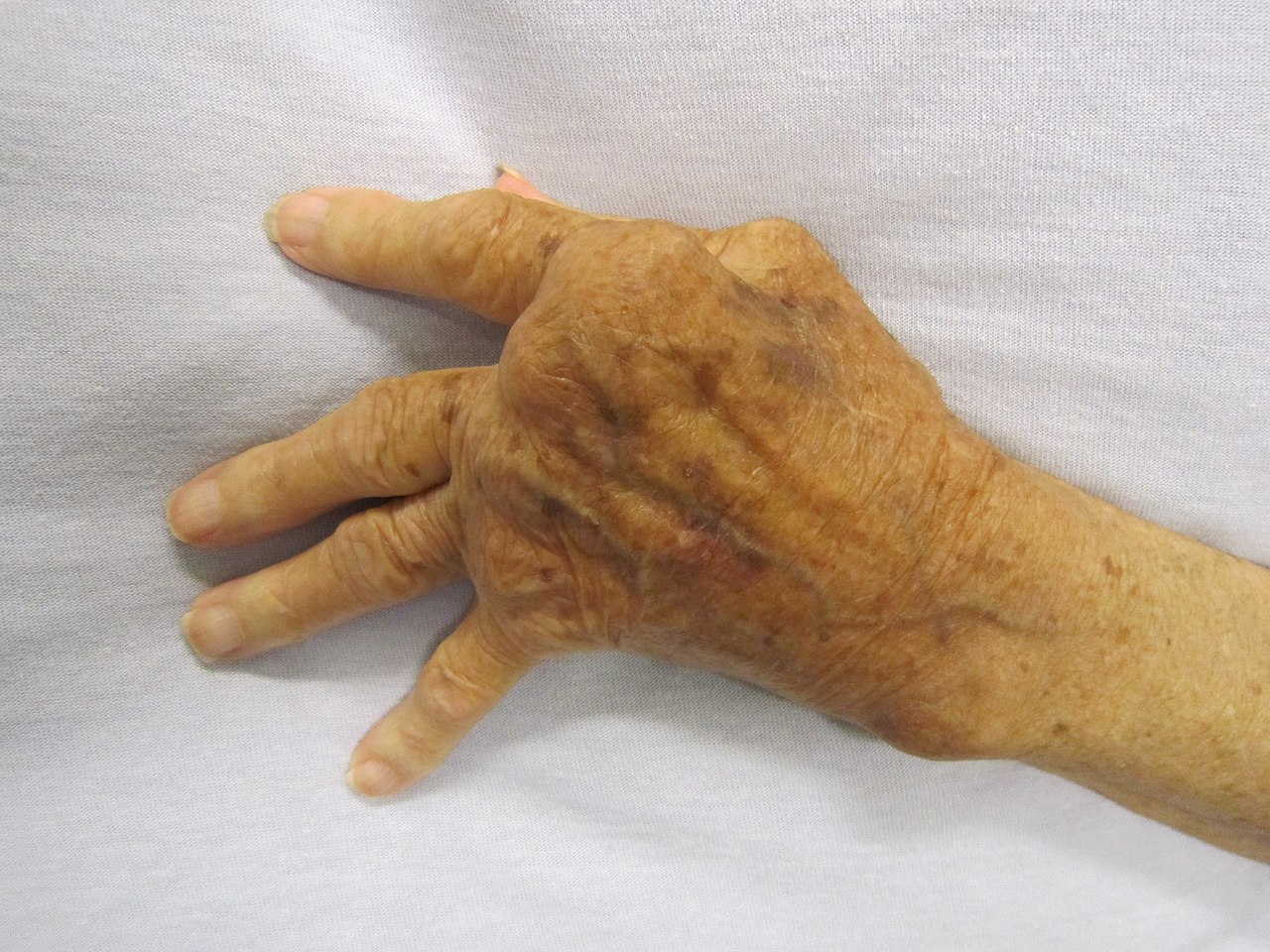
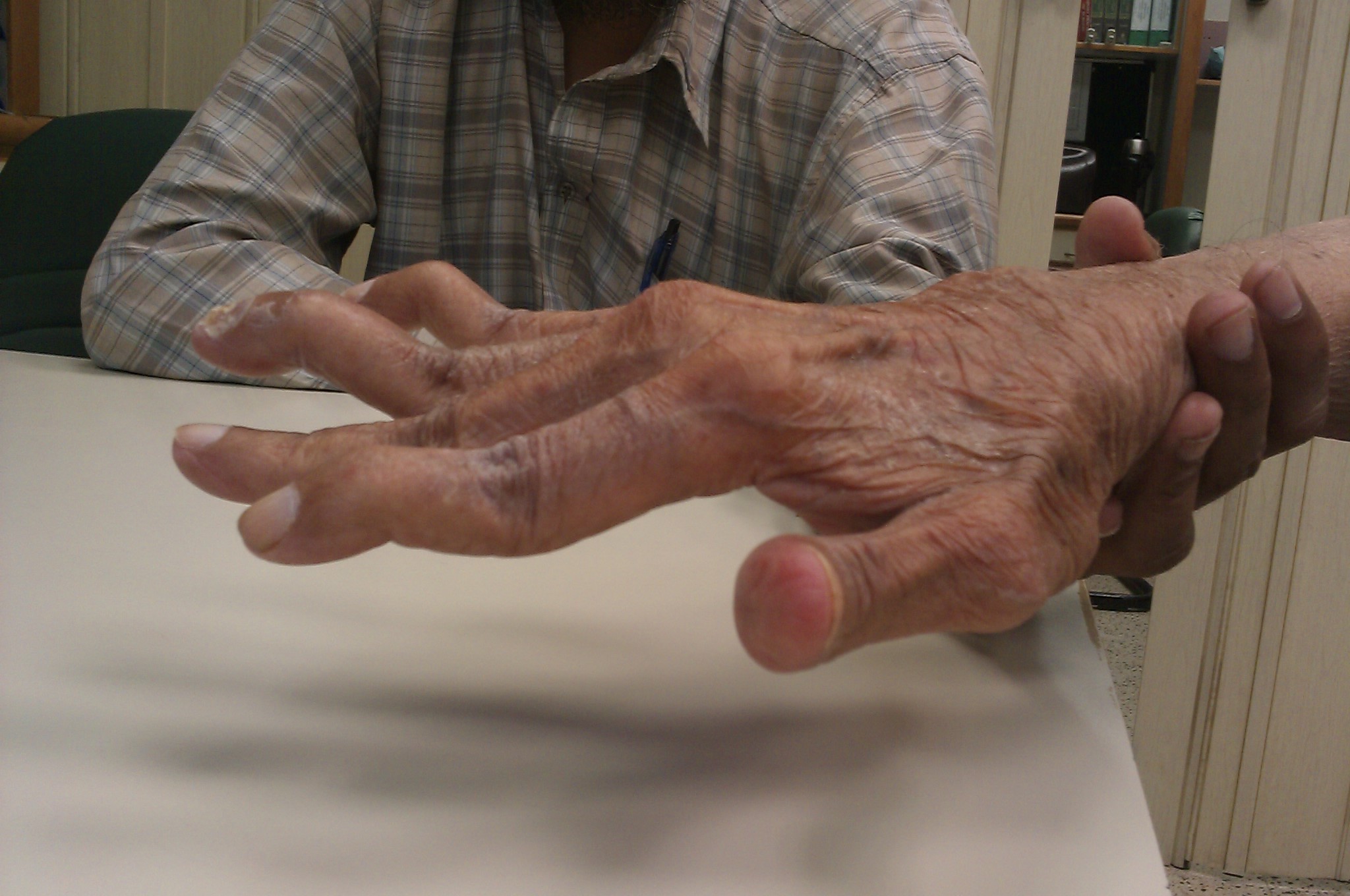
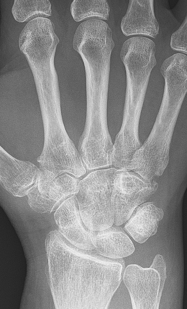
Fig.
9.11 Effects of rheumatoid arthritis on joint structure
https://upload.wikimedia.org/wikipedia/commons/e/ec/Medical_X-Ray_imaging_SKZ07_nevit.jpg
https://live.staticflickr.com/5563/14596721918_63580f8bfb_b.jpg
https://live.staticflickr.com/5558/14596850827_9abd0b72fe_b.jpg
https://upload.wikimedia.org/wikipedia/commons/3/3e/RheumatoideArthritisAP.jpg
Fig.
9.12 Age changes in bone mass
©
Copyright 2020: Augustine G. DiGiovanna, Ph.D., Salisbury University, Maryland
The materials on this site are licensed under CC BY-NC-SA 4.0
![]()
Attribution-NonCommercial-ShareAlike
This license requires that reusers give credit to the
creator. It allows reusers to distribute, remix,
adapt, and build upon the material in any medium or format, for noncommercial
purposes only. If others modify or adapt the material, they must license the
modified material under identical terms.
Previous print editions of the text Human Aging: Biological Perspectives are ©
Copyright 2000, 1994 by The McGraw-Hill Companies, Inc. and 2020 by Augustine
DiGiovanna.
View License Deed | View Legal Code