Some of these figures and pages may be copyright protected. I take no responsibility for matters related to copyright laws and regulations. Use these at your own discretion.
Augustine DiGiovanna, Ph.D.
Fig. 7.1 Image formation and the
pathways used for vision.
Fig. 7.2 Structure of the eye.
https://upload.wikimedia.org/wikipedia/commons/1/15/1413_Structure_of_the_Eye.jpg
https://upload.wikimedia.org/wikipedia/commons/e/e8/Eye-diagram_no_circles_border.svg
Fig. 7.3 Focusing light from (a)
close objects and (b) distant objects.
https://commons.wikimedia.org/wiki/File:Overview_of_the_retina_photoreceptors_(a).png
.png)
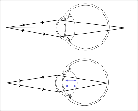
Fig. 7.4 Accommodation to (a)
distant objects and (b) near objects.
https://upload.wikimedia.org/wikipedia/commons/8/82/EyeAccommodation.png
VIDEOS
“Computer-animated
model of accommodation”
Complicated terminology, but good
simple visuals in the video
https://www.youtube.com/watch?v=1yIpyitm6eE
“Accommodation and natural lens”
Simple
clear video but with no audio, only a written explanation.
https://www.dnatube.com/video/4869/Accommodation-and-natural-lens
“The
Near Response of the Eye and Presbyopia, Animation.”
A good video showing the three
changes for near vision – convergence, constriction and accommodation – also
explains presbyopia
https://www.youtube.com/watch?v=ob7laNlslzo
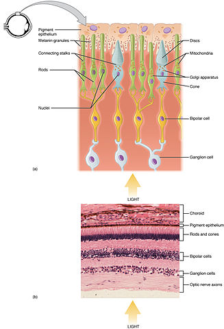
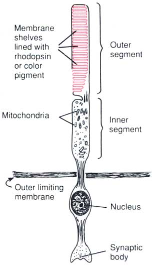
Fig. 7.5 Structure of the retina
and associated eye components.
https://upload.wikimedia.org/wikipedia/commons/b/bb/Rod%26Cone.jpg
https://upload.wikimedia.org/wikipedia/commons/thumb/f/f1/Rod_cell_ca.svg/397px-Rod_cell_ca.svg.png
https://upload.wikimedia.org/wikipedia/commons/2/28/Cone2.svg
https://upload.wikimedia.org/wikipedia/commons/6/6f/Cone_cell.png
Fig. 7.6 The eye in its orbit, with
external eye muscles and fat tissue.
https://upload.wikimedia.org/wikipedia/commons/7/74/1107_The_Extrinsic_Eye_Muscles_right_eye_lat.png
Fig. 7.7 Eyelids and the lacrimal
apparatus.
https://teachmeanatomy.info/wp-content/uploads/The-Lacrimal-Apparatus.jpg
https://www.liberaldictionary.com/wp-content/uploads/2019/02/lacrimal-apparatus-0694.jpg
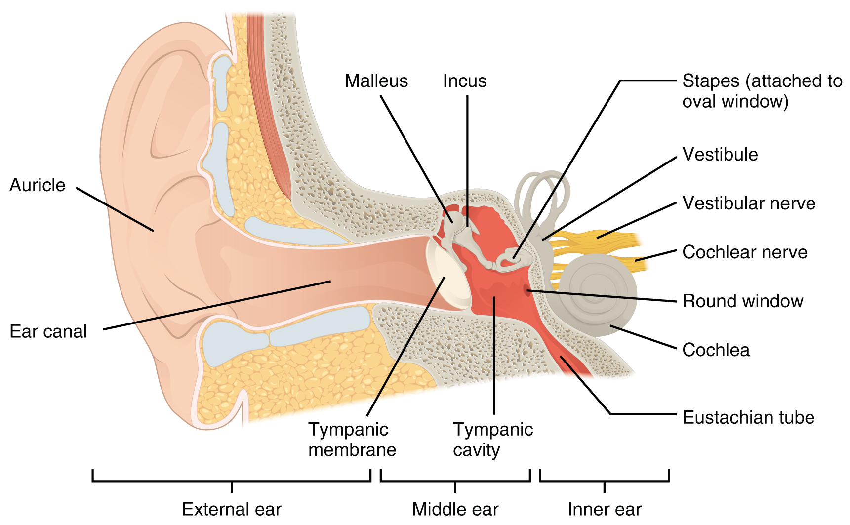
Fig. 7.8
Main structures of the ear.
https://upload.wikimedia.org/wikipedia/commons/4/4b/1404_The_Structures_of_the_Ear.jpg
https://upload.wikimedia.org/wikipedia/commons/c/c4/AnatomyHumanEar.gif
https://search.creativecommons.org/photos/b4809103-bdee-4926-b755-8876dba03108
https://creativecommons.org/licenses/by-sa/3.0/?ref=ccsearch&atype=rich
https://commons.wikimedia.org/wiki/File:Auditory_System.jpg
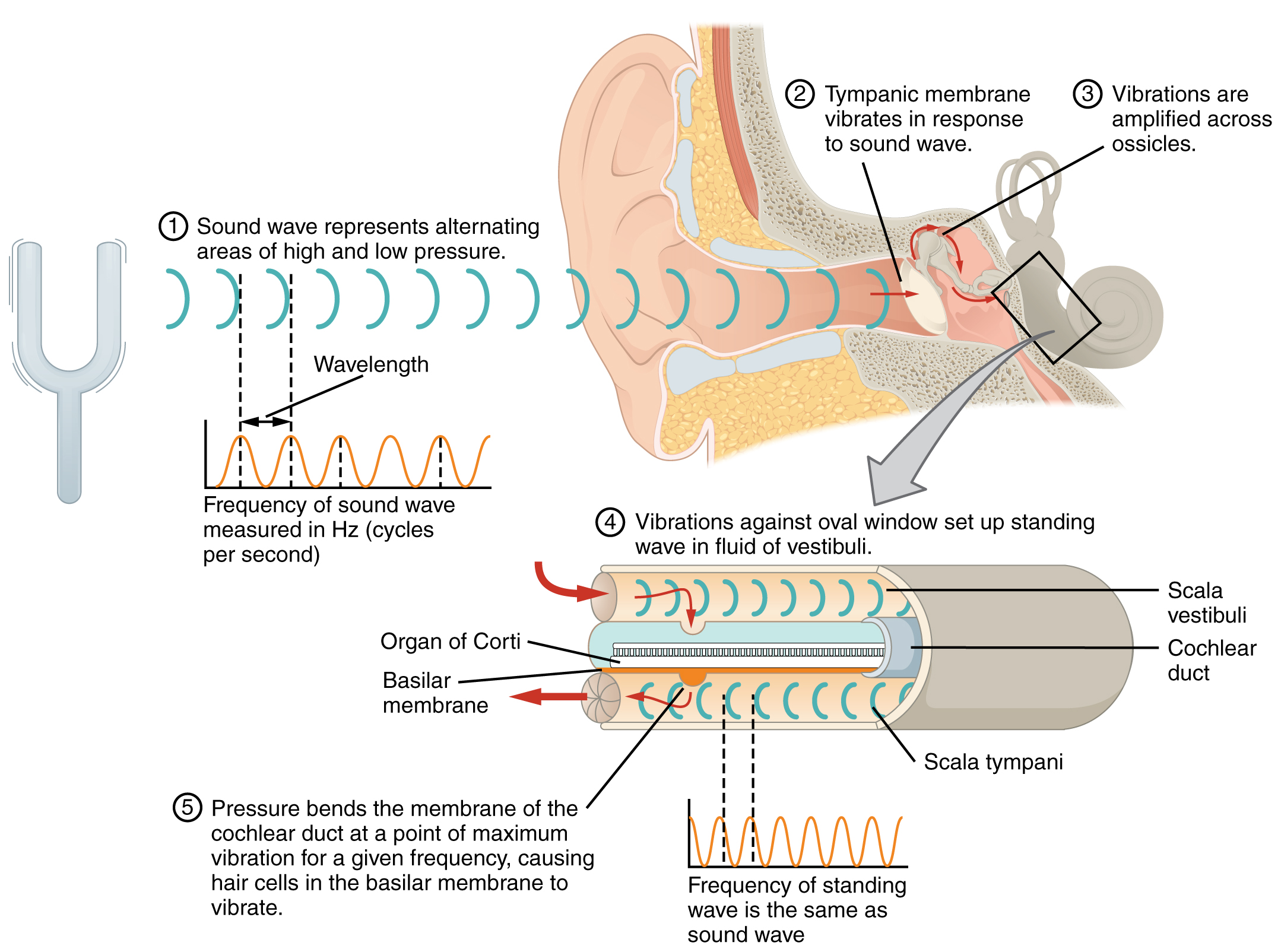
Fig. 7.9 Structures of the middle
ear and the and internal ear
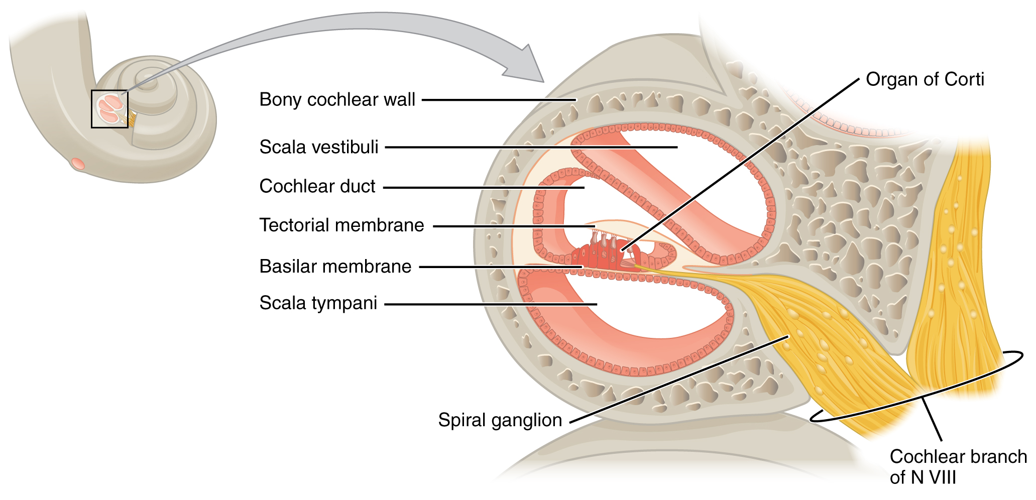
Fig. 7.10 Pathway of sound
vibrations and structure of the cochlea.
https://upload.wikimedia.org/wikipedia/commons/5/51/1405_Sound_Waves_and_the_Ear.jpg
https://upload.wikimedia.org/wikipedia/commons/1/1c/1406_Cochlea.jpg
https://upload.wikimedia.org/wikipedia/commons/0/0c/Cochlea-crosssection.png
https://upload.wikimedia.org/wikipedia/commons/6/6b/1427_Cochlea_Micrograph.jpg
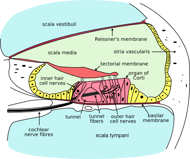
https://upload.wikimedia.org/wikipedia/commons/f/fd/Organ_of_Corti_multilingual.svg
https://upload.wikimedia.org/wikipedia/commons/9/9b/Organ_of_corti.svg
https://commons.wikimedia.org/wiki/File:1407_The_Hair_Cell.jpg
https://commons.wikimedia.org/wiki/File:Hearing_mechanics_cropped.jpg
https://commons.wikimedia.org/wiki/File:1408_Frequency_Coding_in_The_Cochlea.jpg
https://commons.wikimedia.org/wiki/File:L%C3%BBlp%C3%AA%C3%A7_%C3%BB_endam%C3%AA_kort%C3%AE_ku.png
VIDEOS
“The
mechanics of the inner ear”
http://www.animalresearch.info/en/medical-advances/nobel-prizes/the-mechanics-of-the-inner-ear/
Fig. 7.11 Differential cochlear
sensitivities to sound frequencies
Fig. 7.12 The inner ear.
https://upload.wikimedia.org/wikipedia/commons/1/14/Blausen_0329_EarAnatomy_InternalEar.png
VIDEOS
“Hearing
and the cochlea”
https://medlineplus.gov/ency/anatomyvideos/000063.htm
Fig. 7.13 Detecting changes in
gravity and speed
https://upload.wikimedia.org/wikipedia/commons/3/3c/1409_Maculae_and_Equilibrium.jpg
https://commons.wikimedia.org/wiki/File:In%C3%A9rcia_nos_%C3%B3rg%C3%A3os_otol%C3%ADticos.png
https://commons.wikimedia.org/wiki/File:Otolith_organ_of_vestibular_system.jpg
https://commons.wikimedia.org/wiki/File:Balance_Disorder_Illustration_B.png
Fig. 7.14 Detection of rotation
https://upload.wikimedia.org/wikipedia/commons/3/3c/1409_Maculae_and_Equilibrium.jpg
https://commons.wikimedia.org/wiki/File:In%C3%A9rcia_nos_%C3%B3rg%C3%A3os_otol%C3%ADticos.png
https://commons.wikimedia.org/wiki/File:Otolith_organ_of_vestibular_system.jpg
https://commons.wikimedia.org/wiki/File:Balance_Disorder_Illustration_B.png
©
Copyright 2020: Augustine G. DiGiovanna, Ph.D.,
Salisbury University, Maryland
The materials on this site are licensed under CC BY-NC-SA
4.0
![]()
Attribution-NonCommercial-ShareAlike
This license requires that reusers
give credit to the creator. It allows reusers
to distribute, remix, adapt, and build upon the material in any medium
or format, for noncommercial purposes only. If others modify or adapt
the material, they must license the modified material under identical
terms.
Previous print editions of the text Human Aging: Biological Perspectives
are © Copyright 2000, 1994 by The McGraw-Hill Companies, Inc. and 2020
by Augustine DiGiovanna.
View License Deed |
View Legal Code