Some of these figures and pages may be copyright protected.
I take no responsibility for matters related to copyright laws and regulations.
Use these at your own discretion.
Augustine DiGiovanna, Ph.D.
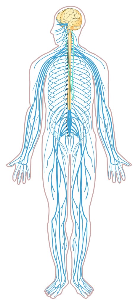
Fig. 6.1 The nervous system.
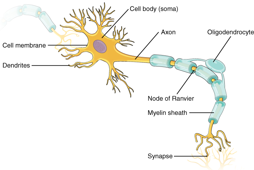
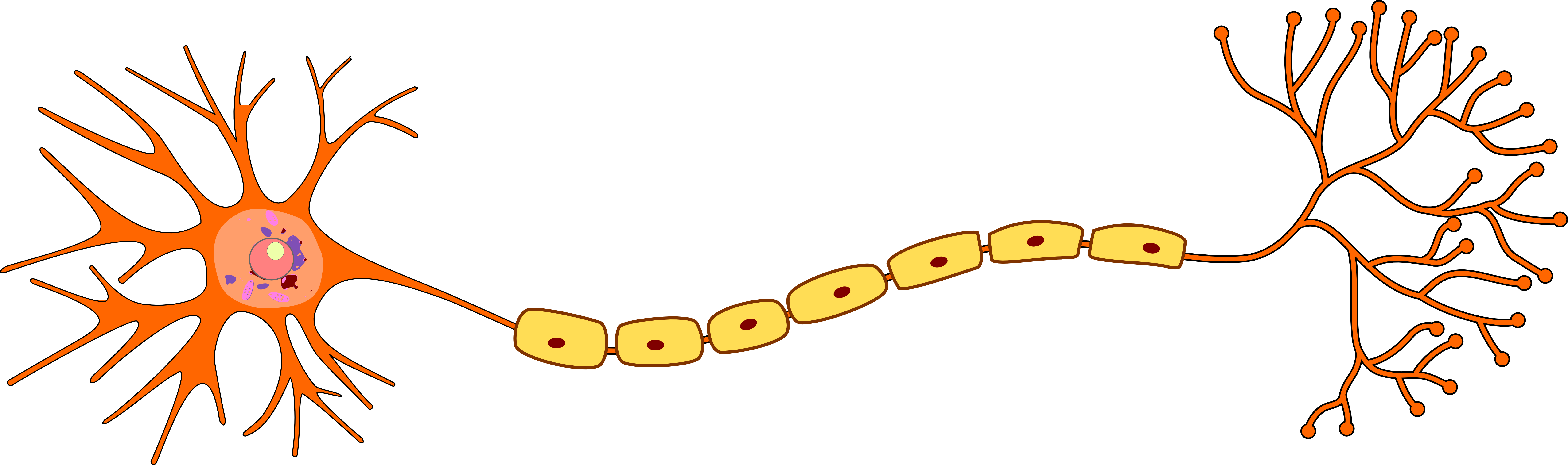
Fig. 6.2 Neuron structure
https://upload.wikimedia.org/wikipedia/commons/6/61/Three_Basic_Types_of_Neuronal_Arrangements.png
https://upload.wikimedia.org/wikipedia/commons/8/86/1206_The_Neuron.jpg
https://upload.wikimedia.org/wikipedia/commons/4/4c/Structure_of_Neuron.png
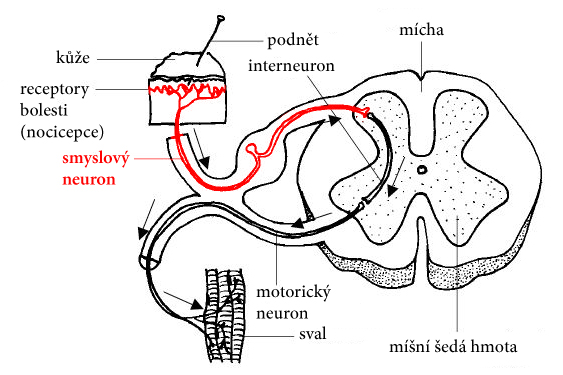
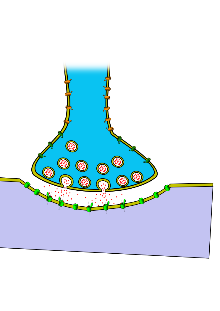
Fig. 6.3 Neuron functioning
https://upload.wikimedia.org/wikipedia/commons/1/1f/Sensory%2C_relay_%26_motor_neurons_cz.jpg
https://upload.wikimedia.org/wikipedia/commons/3/37/1225_Chemical_Synapse.jpg
https://upload.wikimedia.org/wikipedia/commons/d/d7/SynapseSchematic_lines.svg
https://upload.wikimedia.org/wikipedia/commons/d/d1/Synapse_unlabeled.png
https://live.staticflickr.com/2929/14775832941_0745e082e5_b.jpg
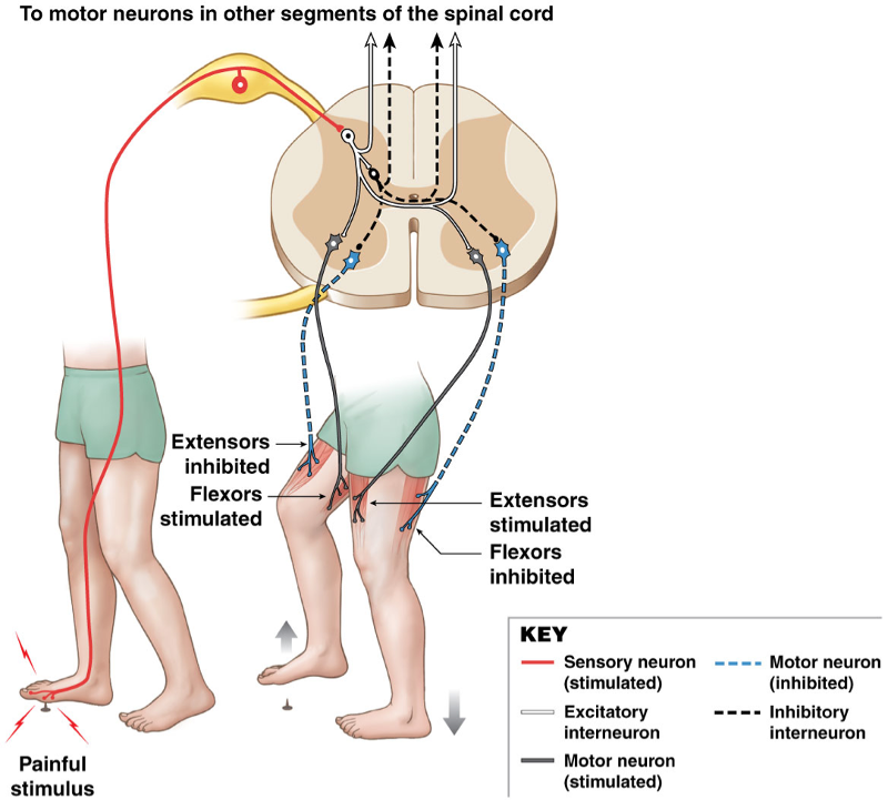
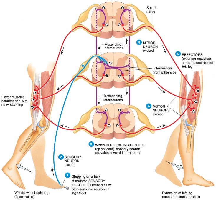
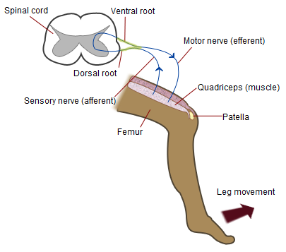
Fig. 6.4
Reflex pathways involving skeletal muscles.
https://www.easynotecards.com/uploads/920/77/1c7a7974_150bb922c9b__8000_00004383.png
https://www.dragonsociety.com/wp-content/uploads/2016/06/crossed_extensor_reflex.jpg
https://upload.wikimedia.org/wikipedia/commons/6/6a/Patellar-knee-reflex.png
https://what-when-how.com/wp-content/uploads/2012/04/tmp14111_thumb2222_thumb.jpg
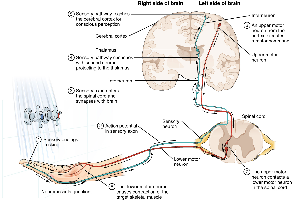
Fig. 6.5 A conscious
sensory pathway.
https://upload.wikimedia.org/wikipedia/commons/6/6e/1212_Sensory_Neuron_Test_Water.jpg
https://upload.wikimedia.org/wikipedia/commons/e/ee/1417_Ascending_Pathways_of_Spinal_Cord.jpg
https://upload.wikimedia.org/wikipedia/commons/e/ee/1417_Ascending_Pathways_of_Spinal_Cord.jpg
https://i.pinimg.com/originals/f2/9c/8f/f29c8fa28363fb40bd23ec4eb59fb67a.jpg
https://o.quizlet.com/5bvtu8EhnEYV49N0sJ.GPg_b.png
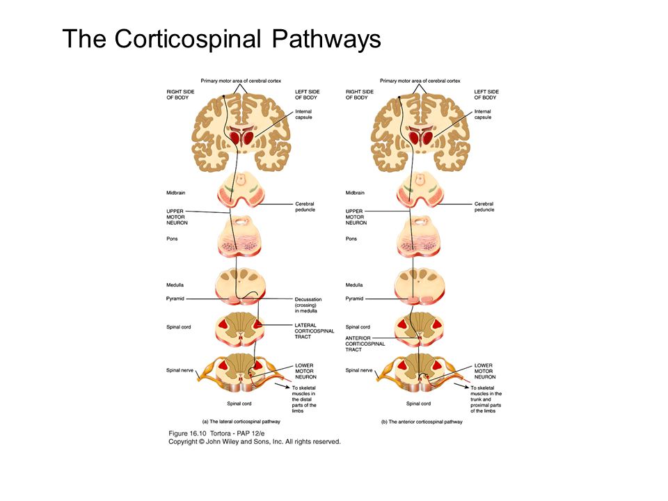
Fig. 6.6 A somatic
(voluntary) motor pathway.
https://upload.wikimedia.org/wikipedia/commons/3/3b/1426_Corticospinal_Pathway.jpg
https://slideplayer.com/slide/10004533/32/images/4/The+Corticospinal+Pathways.jpg
https://o.quizlet.com/aGmWH-nlWIP5aXxadpSL5g.png
Fig. 6.7 Structure of aging brain
https://commons.wikimedia.org/wiki/File:Insula_structure.png
https://commons.wikimedia.org/wiki/File:Right_hemisphere_of_J._Pi%C5%82sudski%27s_brain.jpg
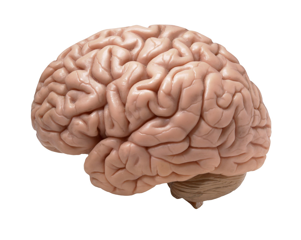
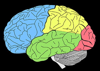
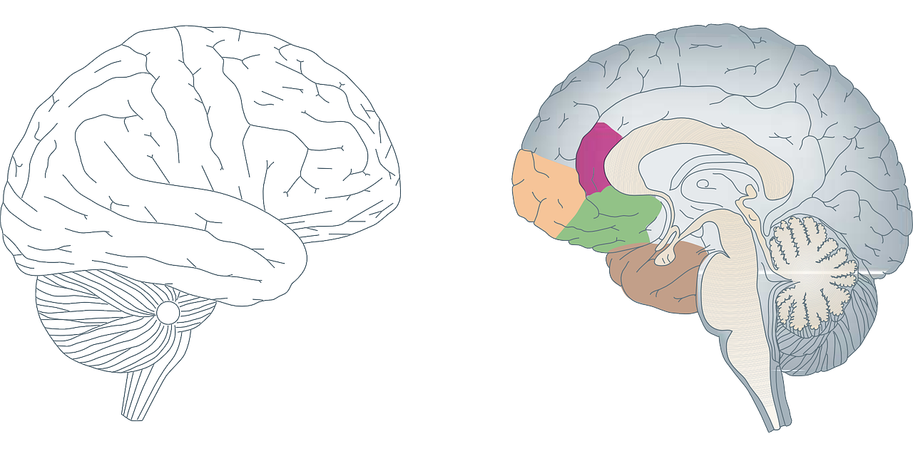
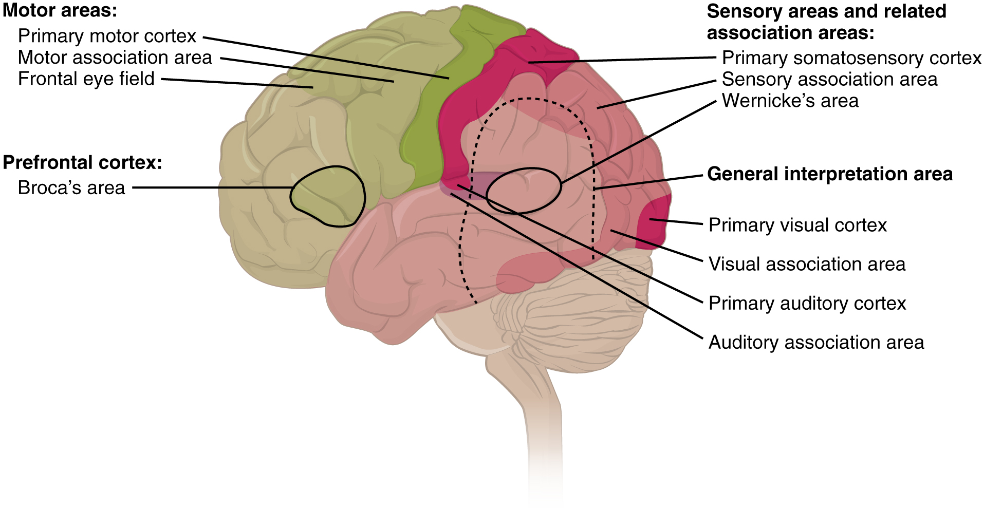
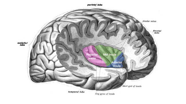
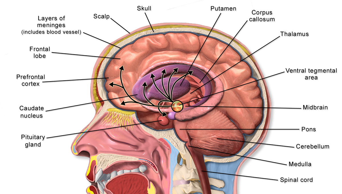
Fig. 6.8 Regions of the brain.
https://upload.wikimedia.org/wikipedia/commons/e/ec/Skull_and_brain_normal_human.svg
https://live.staticflickr.com/8055/8376271918_0ca57957fa_b.jpg
https://live.staticflickr.com/1441/24523085315_51c2ba7beb_b.jpg
https://storage.needpix.com/rsynced_images/brain-150952_1280.png
https://upload.wikimedia.org/wikipedia/commons/8/86/Brain_Anatomy_%28Sagittal%29.png
https://live.staticflickr.com/3521/3824406368_e698492a28_b.jpg
https://upload.wikimedia.org/wikipedia/commons/3/3b/Human_brain_arachnoid_description.JPG
https://upload.wikimedia.org/wikipedia/commons/a/aa/1604_Types_of_Cortical_Areas-02.jpg
https://upload.wikimedia.org/wikipedia/commons/0/00/Insula_structure.png
https://upload.wikimedia.org/wikipedia/commons/2/2b/Brain_Anatomy_Striatum.png
https://upload.wikimedia.org/wikipedia/commons/d/dc/Human_brain.jpg
https://en.wikipedia.org/wiki/File:1311_Brain_Stem.jpg
https://upload.wikimedia.org/wikipedia/commons/thumb/8/8e/Meninges-en.svg/2000px-Meninges-en.svg.png
https://upload.wikimedia.org/wikipedia/commons/a/a4/Active_selection_cognition.png
https://svgsilh.com/svg_v2/2027131.svg
https://upload.wikimedia.org/wikipedia/commons/5/55/Blausen_0076_BasalGanglia.png
https://upload.wikimedia.org/wikipedia/commons/8/85/Basal_ganglia_and_related_structures_%282%29.svg
https://upload.wikimedia.org/wikipedia/commons/7/7d/Brain_bulbar_region.svg
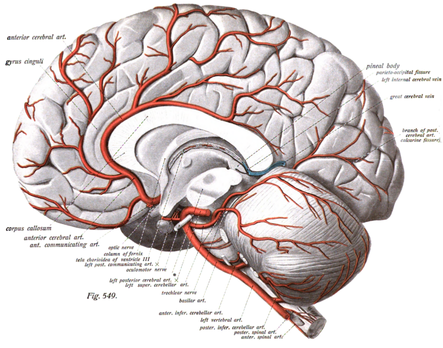
Fig. 6.9 Blood
vessels that supply the brain.
https://upload.wikimedia.org/wikipedia/commons/1/12/2122_Common_Carotid_Artery.jpg
https://upload.wikimedia.org/wikipedia/commons/f/ff/Sobotta.3.1909.549.png
https://upload.wikimedia.org/wikipedia/commons/8/80/Sobo_1909_3_548.png
https://live.staticflickr.com/7752/18007932080_364aaf36f2_b.jpg
https://live.staticflickr.com/5569/14802725833_343efa491a_b.jpg
https://commons.wikimedia.org/wiki/File:Willis_circle_MRI.gif
https://commons.wikimedia.org/wiki/File:Circle_of_willis_from_CT_angio.gif
https://commons.wikimedia.org/wiki/File:2123_Arteries_of_the_Brain.jpg
https://commons.wikimedia.org/wiki/File:Arteries_beneath_brain_Gray_closer.jpg
https://commons.wikimedia.org/wiki/File:Hirnarterien.jpg
https://commons.wikimedia.org/wiki/File:Gray516.png
https://commons.wikimedia.org/wiki/File:Henry_Gray_Figura_516.svg
Fig. 6.10 Strokes
https://commons.wikimedia.org/wiki/File:Two-photon_microscopy_of_in_vivo_brain_function.jpg
https://commons.wikimedia.org/wiki/File:Ischemic_Stroke.svg
https://commons.wikimedia.org/wiki/File:2123_Arteries_of_the_Brain.jpg
https://commons.wikimedia.org/wiki/File:Circle_of_Willis.png
https://commons.wikimedia.org/wiki/File:Stroke_ischemic.jpg
https://commons.wikimedia.org/wiki/File:Cad_anatomy.jpg
https://commons.wikimedia.org/wiki/File:Atrial_fib_stroke.jpg
https://commons.wikimedia.org/wiki/File:Coiled_PCA_residual_aneurysm_arteriogram.JPG
https://commons.wikimedia.org/wiki/File:Sidestream_dark_field_image.jpeg
https://commons.wikimedia.org/wiki/File:Stroke_hemorrhagic.jpg
https://commons.wikimedia.org/wiki/File:Cerebellar_aneurysm.png
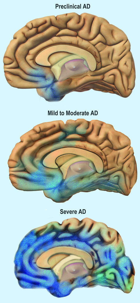
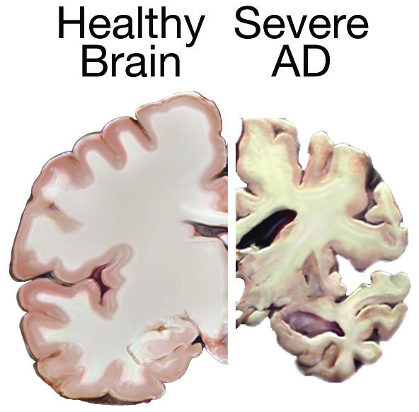
Fig. 6.11 Alzeihmer’s disease
https://live.staticflickr.com/1594/24524716351_f762c70a64_b.jpg
https://live.staticflickr.com/1466/24239522109_80aa4defd1_z.jpg
https://commons.wikimedia.org/wiki/File:Hippocampus_image.png
https://commons.wikimedia.org/wiki/File:Hippocampus_small.gif
https://live.staticflickr.com/4557/38686503251_f0bf300990_b.jpg
https://www.flickr.com/photos/nihgov/38686503251
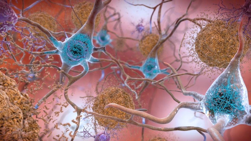
“In the Alzheimer’s affected brain, abnormal levels of the
beta-amyloid protein clump together to form plaques (seen in brown) that
collect between neurons and disrupt cell function. Abnormal collections of the
tau protein accumulate and form tangles (seen in blue) within neurons, harming
synaptic communication between nerve cells.”
Beta-Amyloid Plaques and Tau in the
Brain
https://www.flickr.com/photos/nihgov/25209610730/in/album-72157662951050375/
https://upload.wikimedia.org/wikipedia/commons/9/99/Alzheimers_Disease.jpg
https://upload.wikimedia.org/wikipedia/commons/9/99/Alzheimers_Disease.jpg
https://live.staticflickr.com/1466/24239522109_80aa4defd1_z.jpg
Videos
“Alzheimer disease”
https://medlineplus.gov/ency/anatomyvideos/000003.htm
Fig. 6.12 Basal ganglia
https://commons.wikimedia.org/wiki/File:Basal_ganglia_and_related_structures_(2).svg
Fig. 6.13 Microscopic views of Lewy bodies
https://commons.wikimedia.org/wiki/File:Cuerpos_de_Lewy_y_Neuritas_de_Lewy.png
Copyright 2020: Augustine G. DiGiovanna, Ph.D., Salisbury University, Maryland
The materials on this site are licensed under CC BY-NC-SA
4.0 .
![]()
https://www.biologyofhumanaging.com/Figures/CC-BY-NS-SA%20image.jpg
Attribution-NonCommercial-ShareAlike
This license requires that reusers give
credit to the creator. It allows reusers to distribute, remix, adapt, and build
upon the material in any medium or format, for noncommercial purposes only. If
others modify or adapt the material, they must license the modified material
under identical terms.
Previous print editions of the text Human Aging: Biological Perspectives are ©
Copyright 2000, 1994 by The McGraw-Hill Companies, Inc. and 2020 by Augustine
DiGiovanna.
_(1870)_(14763398095).jpg)
_(14777725651).jpg)