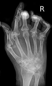
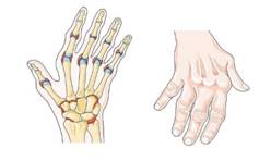
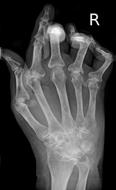
Fig. 9.11 Effects of Rheumatoid arthritis on joint structure. Effects of rheumatoid arthritis on joint structure: (a) Normal joint. (b) Cartilage replaced with pannus. (c) Pannus and immune reaction remove cartilage and bone. (d) Bones fused by calcification of pannus. (Sources of images and videos below. Used with permission.)
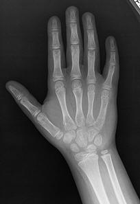
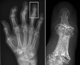

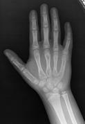
Normal hand RA - Damaged finger joints RA - Fused finger joint


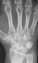
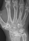
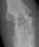
RA – Damaged fingers and wrist RA – Fused wrist bones
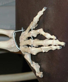
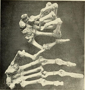
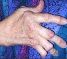
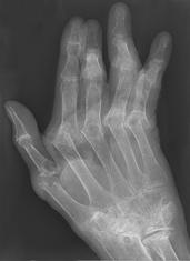
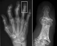
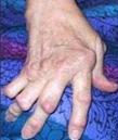
Normal hand bones RA - Damaged hand bones RA - Damaged hand
Videos
“Synovial Membrane”
https://blausen.com/en/video/synovial-membrane/
“Rheumatoid Arthritis – Knee”
https://blausen.com/en/video/rheumatoid-arthritis-knee/
“Rheumatoid Arthritis – Hand”
https://blausen.com/en/video/rheumatoid-arthritis-hand/
©
Copyright 2020: Augustine G. DiGiovanna, Ph.D.,
Salisbury University, Maryland
The materials on this site are licensed under CC BY-NC-SA
4.0
![]()
Attribution-NonCommercial-ShareAlike
This license requires that reusers
give credit to the creator. It allows reusers
to distribute, remix, adapt, and build upon the material in any medium
or format, for noncommercial purposes only. If others modify or adapt
the material, they must license the modified material under identical
terms.
Previous print editions of the text Human Aging: Biological Perspectives
are © Copyright 2000, 1994 by The McGraw-Hill Companies, Inc. and 2020
by Augustine DiGiovanna.
View License Deed |
View Legal Code
Sources of images and videos below. Used
with permission.
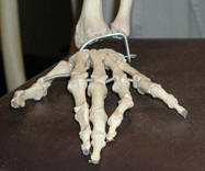
https://commons.wikimedia.org/wiki/File:Hand-bones.jpg
Description English:
Picture of the bones in a human hand (from an authentic human
skeleton). Taken by me on March 25, 2004.
Date 25 March 2004
Source Own work
Author User:Raul654
Permission
(Reusing this file) CC; CC-BY-SA-3.0; Released under the GNU Free Documentation License.
Licensing
I, the copyright holder of this
work, hereby publish it under the following licenses:
Permission is granted to copy,
distribute and/or modify this document under the terms of the GNU Free Documentation
License, Version 1.2 or any later version published by
the Free Software
Foundation; with no Invariant Sections, no Front-Cover
Texts, and no Back-Cover Texts. A copy of the license is included in the
section entitled GNU Free Documentation License.
This file is licensed under the Creative commons Attribution-Share Alike 3.0 Unported license.
You are free:
·
to share – to copy, distribute and transmit the work
·
to remix – to adapt the work
Under the following conditions:
·
attribution – You must give appropriate credit, provide a link to the license, and
indicate if changes were made. You may do so in any reasonable manner, but not
in any way that suggests the licensor endorses you or your use.
share alike – If you remix, transform, or build upon the
material, you must distribute your contributions under the same or compatible license as the original.
You may select the license of
your choice.
https://commons.wikimedia.org/wiki/File:Rheumatoid_arthritis_--_Smart-Servier_(cropped).jpg
.jpg)
Author Laboratoires Servier
Title rheumatoid
arthritis
Object type designated
intractable/rare diseases ![]()
Description English: Skeleton and bones - Rheumatoid arthritis
Date 29 September 2019
·
Authority control : Q187255
·
Source/Photographer Smart Servier
website: Images related to Rheumatoid
arthritis, Skeleton and bones and Bones -- Download in Powerpoint format.
Flickr: Images related to
Rheumatoid arthritis, Skeleton and bones and Bones (in French).
Other versions This file has been extracted
from another file: Rheumatoid arthritis -- Smart-Servier.jpg
Related images
Licensing
This is an freely
reusable image from SMART-Servier Medical Art, part of Laboratoires Servier.
This tag does not
indicate the copyright status of the attached work. A normal copyright tag is still required. See commons:Licensing.
This file is licensed under the Creative commons Attribution-Share Alike 3.0 Unported license.
You are free:
·
to share – to copy, distribute and transmit the work
·
to remix – to adapt the work
Under the following
conditions:
·
attribution – You must give appropriate credit, provide a link to the license, and
indicate if changes were made. You may do so in any reasonable manner, but not
in any way that suggests the licensor endorses you or your use.
share alike – If you remix, transform, or build upon the
material, you must distribute your contributions under the same or compatible license as the original.
https://commons.wikimedia.org/wiki/File:Rheumatoid_arthritis_with_unaffected_carpal_bones_2009.jpg
|
Description |
English: X-ray of the wrist of a then 58 year old
woman with rheumatoid
arthritis, showing unaffected carpal bones. 8 years later,
she had developed ankylosing fusion of the bones - see File:Rheumatoid arthritis with carpal ankylosis
2017.jpg. |
|
Date |
19 January 2009 |
|
Source |
Own work |
|
Author |
Licensing
I, the copyright holder
of this work, hereby publish it under the following license:
This file is made available under the Creative commons CC0 1.0 Universal Public Domain Dedication.
The person who associated a work with this deed has dedicated the work to
the public domain by waiving all of their rights to the work worldwide under copyright law,
including all related and neighboring rights, to the extent allowed by law. You
can copy, modify, distribute and perform the work, even for commercial
purposes, all without asking permission.
https://commons.wikimedia.org/wiki/File:Rheumatoid_arthritis_with_carpal_ankylosis_2017.jpg
Description English:
X-ray of the wrist of a 66 year old
woman with rheumatoid arthritis, showing ankylosing fusion of the carpal bones. Previous X-ray showed unaffected carpal bones - see File:Rheumatoid arthritis with unaffected carpal bones 2009.jpg.
Date 19 January 2017
Source Own work
Author Mikael Häggström
Licensing
I, the copyright holder
of this work, hereby publish it under the following license:
This file is made available under the Creative commons CC0 1.0 Universal Public Domain Dedication.
The person who associated a work with this deed has dedicated the work to
the public domain by waiving all of their rights to the work worldwide under copyright law,
including all related and neighboring rights, to the extent allowed by law. You
can copy, modify, distribute and perform the work, even for commercial
purposes, all without asking permission.
Description English: Projectional radiography ("X-ray") of a normal hand of a 8 year
old male, by dorsoplantar view.
Date 14 March
2018
Source bonepit.com
Author Staff at the
Department of Radiology, UC San Diego Health.
Permission
(Reusing this file) See below.
Licensing
This file is in the public domain because it is a
work of medical imaging created in the United States and does not contain additional
copyrightable graphics. See Meta:wikilegal/Copyright of Medical Imaging for details.
Informed consent is generally required at least where human subjects are identifiable.
This work is free and may be used by anyone for any purpose. If you wish to use this content, you do not need to
request permission as long as you follow any licensing requirements mentioned
on this page.
wikimedia
Foundation has received an e-mail confirming that the copyright holder has
approved publication under the terms mentioned on this page. This
correspondence has been reviewed by
an OTRS member and stored in our permission
archive. The correspondence is available to trusted
volunteers ticket
#2018030210000886.
If you have questions about the
archived correspondence, please use the OTRS noticeboard. Ticket link: https://ticket.wikimedia.org/otrs/index.pl?Action=AgentTicketZoom&TicketNumber=2018030210000886
https://commons.wikimedia.org/wiki/File:RheumatoideArthritisAP.jpg
Description Typisches Röntgenbild einer
Rheumatoiden Arthritis.
Date Unknown date
Source Own work
Author Bernd Brägelmann Braegel Mit freundlicher Genehmigung von Dr. Martin Steinhoff
Licensing
I, the copyright holder of this
work, hereby publish it under the following licenses:
Permission is
granted to copy, distribute and/or modify this document under the terms of the GNU Free
Documentation License, Version 1.2 or any
later version published by the Free Software
Foundation; with no Invariant Sections, no Front-Cover
Texts, and no Back-Cover Texts. A copy of the license is included in the
section entitled GNU Free Documentation License.
This file is licensed under the Creative commons Attribution 3.0 Unported license.
You are free:
·
to share – to copy, distribute and transmit the work
·
to remix – to adapt the work
Under the following
conditions:
attribution – You must give appropriate credit, provide a
link to the license, and indicate if changes were made. You may do so in any
reasonable manner, but not in any way that suggests the licensor endorses you
or your use.
You may select the license of
your choice.
Description English:
X-ray of right fourth proximal
interphalangeal (PIP) joint with bone erosions by rheumatoid arthritis. Taken October 2002. Same joint is partially healed on a follow-up X-ray
after treatment with conventional disease-modifying
antirheumatic drugs(DMARDs) one year later: File:X-ray of right fourth PIP joint with partially healed bone erosions by
rheumatoid arthritis.jpg
Date 21 March 2006
Source (2006). "Bone erosions in rheumatoid arthritis can be
repaired through reduction in disease activity with conventional
disease-modifying antirheumatic drugs". Arthritis
Research & Therapy 8
(3): R76. DOI:10.1186/ar1943.
ISSN 14786354. (CC-BY-2.0)
Author Haruko Ideguchi, Shigeru Ohno, Hideaki Hattori, Akiko Senuma and Yoshiaki Ishigatsubo
Licensing
This file is licensed under the Creative commons Attribution 2.0 Generic license.
You are free:
·
to share – to copy, distribute and transmit the work
·
to remix – to adapt the work
Under the following
conditions:
attribution – You must give appropriate credit, provide a link
to the license, and indicate if changes were made. You may do so in any
reasonable manner, but not in any way that suggests the licensor endorses you
or your use.
https://commons.wikimedia.org/wiki/File:Rheumatoide_Arthritis_der_Hand_65W_-_CR_ap_-_001.jpg
Description Deutsch: Rheumatoide Arthritis der Hand. Zusätzlich Fingerfrakturen.
Date 10 December 2020
Source Own work
Author Hellerhoff
Licensing
I, the copyright
holder of this work, hereby publish it under the following license:
This file is licensed under the Creative commons Attribution-Share Alike 4.0 International license.
You are free:
·
to share – to copy, distribute and transmit the work
·
to remix – to adapt the work
Under the following
conditions:
·
attribution – You must give appropriate credit, provide a link to the license, and indicate
if changes were made. You may do so in any reasonable manner, but not in any
way that suggests the licensor endorses you or your use.
share alike – If you remix, transform, or build upon the
material, you must distribute your contributions under the same or compatible license as the original.
_(14760829644).jpg)
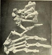
Description English:
Hands: Arthritis deformans (rheumatoid arthritis)
Identifier:
bulletinofwarren00harv (find
matches)
Title: Bulletin of
the Warren Anatomical Museum
Year: 1910 (1910s)
Authors: Harvard
Medical School Whitney,
William F
Subjects: Warren
Anatomical Museum Anatomy,
Pathological Museums Anatomy Pathology
Publisher: Boston :
(Harvard Medical School)
Contributing Library: Francis
A. Countway Library of Medicine
Digitizing Sponsor: Open
Knowledge commons and Harvard Medical School
View Book Page: Book
Viewer
About This Book: Catalog Entry
View All Images: All
Images From Book
Click here to view book
online to see this illustration in context in a browseable online version of this book.
Text Appearing Before Image:
efingers point downward toward the palm,
directed toward the cubital edge ofthe hand. There is
growth of bone about the joint, with subluxation of thephalanges
and atrophy of the bones. From an adult. 1874. Henry W. Dean. F. W. Stackpole.
I359« Finger. The bones of the finger in connection, dried.They show a strong lateral inclination of the
terminal phalanx upon thesecond, but without any
appearance of disease. 1859. Dr. R. M. Hodges. 6035. Finger. Anchylosis. The
terminal phalanges of two fingersThe second phalanx
is dislocated upward and forward on the bone in both cases and anchylosed in a
new position. In one the two bones form an obtuse angle, and in the other a
right angle. From a woman about 75 years old. Twenty years before she had bed
sores upon the hand and suffered from them for about two years. 1849. Dr. J. B.
S. Jackson. 6377. Finger. The phalanges of the finger, dried. The last two
anchylosed with formation of new bone about the joint. JOINTS.—ARTHRITIS
DEFORMANS. 53
Text Appearing After Image:
4305. Hand. Arthritis Deiormans. 10207. Hands.
Both hands articulated and dried. There is an extensive formation of new bone
about the joints of theterminal phalanges which must
have greatly impeded their motion. There isalso a
similar deposit but less extensive at the articulation of the thumb withthe wrist. Dr. Thomas Dwight. 10206. Foot. An
articulated foot, dried. There are numerous irregular osseous growths over the
surface, especiallyin the neighborhood of the
phalangeal articulation. From a man 45 years old Dr. Thomas Dwight. 54 JOINTS.—ARTHRITIS DEFORMANS. 4744. Astragalus. The
astragalus, dried. There is a piece of imperfectly developed bone attached to
the upper edgeof the articular surface. The joint
surface is eburnated and greasy. 1876. 1366.
Astragalus. Os Calcis. The
astragalus and os calcis, dried.They are partially, but
strongly anchylosed, with growths of new boneabout,
the edges of the articular surfaces. 1847, Dr. J. C. Warren. 5134. Sacrum,
dried. There is stron
Note About Images
Please note that these images are extracted from scanned page images that
may have been digitally enhanced for readability - coloration and appearance of
these illustrations may not perfectly resemble the original work.
Date 1910
Source https://www.flickr.com/photos/internetarchivebookimages/14760829644/
·
Source book page: https://archive.org/stream/bulletinofwarren00harv/bulletinofwarren00harv#page/n64/mode/1up
Author Internet Archive Book Images
Permission
(Reusing this file) At the time of upload, the image license
was automatically confirmed using the Flickr API. For more information see Flickr API detail.
Flickr posted date 28 July 2014
Licensing
This image was taken
from Flickr's The commons. The uploading organization
may have various reasons for determining that no known copyright
restrictions exist, such as:
1.
The copyright is in
the public domain because it has expired;
2.
The copyright was
injected into the public domain for other reasons, such as failure to adhere to
required formalities or conditions;
3.
The institution owns
the copyright but is not interested in exercising control; or
4.
The institution has
legal rights sufficient to authorize others to use the work without
restrictions.
More
information can be found at https://flickr.com/commons/usage/.
Please add
additional copyright tags to this image if more specific information about
copyright status can be determined. See commons:Licensing for more information.
This image was
originally posted to Flickr by Internet Archive Book Images at https://flickr.com/photos/126377022@N07/14760829644. It was reviewed on 23 October 2015 by FlickreviewR and was confirmed to be licensed under the terms of the No known copyright
restrictions.
https://commons.wikimedia.org/wiki/File:Erosive_osteoarthritis_with_gull-wing_appearance.jpg
Description English:
X-ray of the hand of a 76-year old female farmer. There
were no symptoms or blood tests indicating rheumatoid arthritis or gout,
conferring a diagnosis of osteoarthritis. The X-ray shows a gull-wing appearance of
involved joints, thus being erosive osteoarthritis.
Date 3 December 2018
Source Own work
Author
Mikael Häggström, M.D.
- Author info
- Reusing images
With seagull
Licensing
This file is made available under the Creative commons CC0 1.0 Universal Public Domain Dedication.
The person who associated a work with this deed has dedicated the work to
the public domain by waiving all of their rights to the work worldwide under copyright law,
including all related and neighboring rights, to the extent allowed by law. You
can copy, modify, distribute and perform the work, even for commercial
purposes, all without asking permission.
https://commons.wikimedia.org/wiki/File:Arthrite_rhumatoide_late.jpg
Description English:
Čeština: Revmatoidní
artritida - postižení kloubů ruky
English: Rheumatoid arthritis
- affected joints of the hand
Français : Arthrite rhumatoide - articulations touchées de la main
Date 15 August 2007,
22:52:56
·
Source Original source: http://nihseniorhealth.gov/arthritis/toc.html
·
Originally from fr.wikipedia; description page is/was here.
Based off: File:Arthrite rhumatoide.jpg
Author NIHSeniorhealth
Licensing
This work is in the public domain in the United
States because it is a work prepared by an officer or employee of the United States Government as
part of that person’s official duties under the terms of Title 17, Chapter 1, Section 105 of the US Code. Note: This only applies to original works of the Federal Government and not to
the work of any individual U.S. state, territory, commonwealth, county, municipality, or any other subdivision. This
template also does not apply to postage stamp designs published by the United States Postal
Service since 1978. (See § 313.6(C)(1) of Compendium of
U.S. Copyright Office Practices). It also does not apply to certain US coins;
see The US Mint Terms of Use.