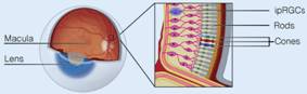
Fig. 7.5a Structure of the retina and associated eye components. (Scroll down for cone and rod.) (Sources of images and videos below. Used with permission.)
Videos
“The Eye”
https://blausen.com/en/video/the-eye/
“The Posterior Cavity”
https://blausen.com/en/video/the-posterior-cavity/
“The Rretina”
https://blausen.com/en/video/the-retina/
“The Optic Disc Blind Spot”
https://blausen.com/en/video/the-optic-disc-blind-spot/
“The Fovea of the Retina”
https://blausen.com/en/video/the-fovea-of-the-retina/
“The Vascular Tunic”
https://blausen.com/en/video/the-vascular-tunic/
©
Copyright 2020: Augustine G. DiGiovanna, Ph.D.,
Salisbury University, Maryland
The materials on this site are licensed under CC BY-NC-SA
4.0
![]()
Attribution-NonCommercial-ShareAlike
This license requires that reusers
give credit to the creator. It allows reusers
to distribute, remix, adapt, and build upon the material in any medium
or format, for noncommercial purposes only. If others modify or adapt
the material, they must license the modified material under identical
terms.
Previous print editions of the text Human Aging: Biological Perspectives
are © Copyright 2000, 1994 by The McGraw-Hill Companies, Inc. and 2020
by Augustine DiGiovanna.
View License Deed |
View Legal Code
Sources of images and videos. Used
with permission.
https://commons.wikimedia.org/wiki/File:Overview_of_the_retina_photoreceptors_(a).png
.png)
Description English: Overview of the retina photoreceptors.
Schematic view of the eye with the retina at the back of the eye (the fundus),
containing cones, rods and the intrinsically photosensitive retinal ganglion
cells (ipRGCs) expressing the photopigment melanopsin.
Date
Published: 20 August 2019
Source
Blume, C.,
Garbazza, C. & Spitschan, M. Effects of light
on human circadian rhythms, sleep and mood. Somnologie 23, 147–156 (2019).
https://doi.org/10.1007/s11818-019-00215-x
Author
Christine Blume, Corrado
Garbazza
& Manuel Spitschan
Licensing
This file is
licensed under the
Creative commons
Attribution 4.0 International license.
You are free:
·
to share – to
copy, distribute and transmit the work
·
to remix – to
adapt the work
Under the following conditions:
attribution – You must give
appropriate credit, provide a link to the license, and indicate if changes were
made. You may do so in any reasonable manner, but not in any way that suggests
the licensor endorses you or your use.
https://commons.wikimedia.org/wiki/File:Cone2.svg
Description English:
Rod cell
Source Own work
Author
Madhero88
Other versions Derivative works of this file:
.svg)
Licensing
I, the copyright holder of this work, hereby publish it
under the following license:
This file is licensed under the
Creative commons
Attribution-Share Alike 3.0 Unported license.
You are free:
·
to share – to copy, distribute and
transmit the work
·
to remix – to adapt the work
Under the following conditions:
·
attribution – You must give appropriate
credit, provide a link to the license, and indicate if changes were made. You
may do so in any reasonable manner, but not in any way that suggests the
licensor endorses you or your use.
share alike – If you remix, transform, or build upon the material,
you must distribute your contributions under the
same or compatible license as the original.
Modified:
A.G.DiGiovanna
Added descriptions, items, labels,
and lines.
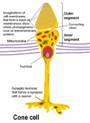
https://commons.wikimedia.org/wiki/File:Cone_cell_en.png
Description English:
Cone cell.
Eesti:
Kolvikese joonis
Date
18 March 2010
Source Own work
Author
Ivo Kruusamägi
Permission
(Reusing this file)
When using this picture in non-wikipedia
site the name of the author and the name wikipedia (or
Vikipeedia) should be mentioned.
Other versions
This
image is based on my own made
Cone cell.png that
has text in Estonian.
Licensing
I, the copyright holder of this work, hereby publish it
under the following license:
This file is licensed under the
Creative commons
Attribution-Share Alike 3.0 Unported license.
You are free:
·
to share – to copy, distribute and
transmit the work
·
to remix – to adapt the work
Under the following conditions:
·
attribution – You must give appropriate
credit, provide a link to the license, and indicate if changes were made. You
may do so in any reasonable manner, but not in any way that suggests the
licensor endorses you or your use.
share alike – If you remix, transform, or build upon the material,
you must distribute your contributions under the
same or compatible license as the original.
Videos
“The Eye”
https://blausen.com/en/video/the-eye/
“The Posterior Cavity”
https://blausen.com/en/video/the-posterior-cavity/
“The Rretina”
https://blausen.com/en/video/the-retina/
“The Optic Disc Blind Spot”
https://blausen.com/en/video/the-optic-disc-blind-spot/
“The Fovea of the Retina”
https://blausen.com/en/video/the-fovea-of-the-retina/
“The Vascular Tunic”
https://blausen.com/en/video/the-vascular-tunic/