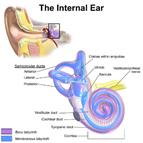
Fig. 7.12 The inner ear. (Sources of
images and videos below. Used with permission.)
Videos
“Internal Ear Anatomy”
https://blausen.com/en/video/internal-ear-anatomy/
https://www.biologyofhumanaging.com/Figures/CC-BY-NS-SA%20image.jpg
Attribution-NonCommercial-ShareAlike
This
license requires that reusers give credit to the
creator. It allows reusers to distribute, remix,
adapt, and build upon the material in any medium or format, for noncommercial
purposes only. If others modify or adapt the material, they must license the
modified material under identical terms.
Previous print editions of the text Human Aging: Biological Perspectives are ©
Copyright 2000, 1994 by The McGraw-Hill Companies, Inc. and 2020 by Augustine
DiGiovanna.
View License Deed | View Legal Code
Sources of images and videos below. Used with
permission.
https://commons.wikimedia.org/wiki/File:Blausen_0329_EarAnatomy_InternalEar.png
Description English: Internal
Ear Anatomy. See a related animation of this medical topic.
Date 15 October 2013, 14:40:06
Source Own work
Author BruceBlaus. When using this image in external sources it can be cited as:
·
Blausen.com staff (2014). "Medical gallery of Blausen Medical 2014". wikiJournal of Medicine 1 (2). DOI:10.15347/wjm/2014.010. ISSN 2002-4436.
Permission
(Reusing this file) This work is free and may be used by anyone for any purpose. If you wish to use this content, you do not need to
request permission as long as you follow any licensing requirements mentioned
on this page.
wikimedia Foundation has received an e-mail confirming
that the copyright holder has approved publication under the terms mentioned on
this page. This correspondence has been reviewed
by an OTRS
member and stored in our permission archive. The
correspondence is available to trusted volunteers ticket #2013061010006654.
If you have questions about the archived correspondence,
please use the OTRS
noticeboard. Ticket link: https://ticket.wikimedia.org/otrs/index.pl?Action=AgentTicketZoom&TicketNumber=2013061010006654
This work is free and may be used by
anyone for any purpose. If you wish to use this content, you do not need to
request permission as long as you follow any licensing requirements mentioned
on this page.
wikimedia Foundation has received an e-mail confirming
that the copyright holder has approved publication under the terms mentioned on
this page. This correspondence has been reviewed
by an OTRS
member and stored in our permission archive. The
correspondence is available to trusted volunteers ticket #2013061010006654
Modified: A.G. DiGiovanna
Added labels and
lines. Blocked some portions.
https://commons.wikimedia.org/wiki/File:Vestibular_organs-_canals,_otolith,_cochlea.jpg
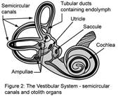
Description a drawing of the inner ear
Source The Effects of Space Flight on the Human Vestibular System, an online educational article by the
U.S. government’s National Aeronautics and Space Administration (NASA)
Author NASA
Licensing
This file is in the public domain in the United States because it was solely
created by NASA. NASA copyright policy
states that "NASA material is not protected by copyright unless noted".
(See Template:PD-USGov, NASA copyright policy page or JPL Image Use Policy.)
Warnings:
- Use of NASA logos, insignia and
emblems is restricted per U.S. law 14 CFR 1221.
- The NASA website hosts a large number of images from
the Soviet/Russian space agency, and other
non-American space agencies. These are not necessarily in the
public domain.
- Materials based on Hubble Space Telescope data may be copyrighted if they are not explicitly
produced by the STScI.[1] See also {{PD-Hubble}} and {{Cc-Hubble}}.
- The SOHO (ESA &
NASA) joint project implies
that all materials created by its probe are copyrighted and require
permission for commercial non-educational use. [2]
- Images featured on the Astronomy Picture of
the Day (APOD) web
site may be copyrighted. [3]
The National Space Science Data Center (NSSDC) site has been known to host copyrighted content. Its photo gallery FAQ states that all of
the images in the photo gallery are in the public domain "Unless otherwise
noted."
Modified: A.G. DiGiovanna
Added labels and
lines. Blocked some portions.
https://commons.wikimedia.org/wiki/File:Ampulla_of_SemicircularCanal.svg
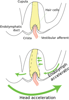
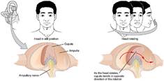
Description English: The
cupula of the human semicircular canal. Top: The cupula spans the lumen of the ampulla
from the crista to the membranous labyrinth. Bottom: Since head acceleration
exceeds endolymph acceleration, the relative flow of endolymph in the canal is
opposite to the direction of head acceleration. This flow produces a pressure
across the elastic cupula, which deflects in response.
Date 16 February 2011
Source Own work
Author Thomas.haslwanter
Licensing
I, the copyright holder of this work, hereby publish it
under the following licenses:
This file is
licensed under the Creative commons Attribution-Share Alike 3.0 Unported license.
You are free:
·
to share – to copy,
distribute and transmit the work
·
to remix – to adapt the work
Under the following conditions:
·
attribution – You must give
appropriate credit, provide a link to the license, and indicate if changes were
made. You may do so in any reasonable manner, but not in any way that suggests
the licensor endorses you or your use.
share alike – If you remix,
transform, or build upon the material, you must distribute your contributions
under the same or compatible license as the original.
Permission is
granted to copy, distribute and/or modify this document under the terms of the GNU Free Documentation License, Version 1.2 or any
later version published by the Free Software Foundation; with no Invariant Sections, no Front-Cover
Texts, and no Back-Cover Texts. A copy of the license is included in the
section entitled GNU Free Documentation License.
You may select the license of your choice.
Modified: A.G. DiGiovanna
Added labels and
lines. Blocked some portions.
https://commons.wikimedia.org/wiki/File:1410_Equilibrium_and_Semicircular_Canals.jpg
Description English: Illustration
from Anatomy & Physiology, Connexions Web site. http://cnx.org/content/col11496/1.6/, Jun 19, 2013.
Date 28 May 2013, 00:56:05
Source Anatomy &
Physiology, Connexions Web site. http://cnx.org/content/col11496/1.6/, Jun 19, 2013.
Author OpenStax College
Licensing
This file is
licensed under the Creative commons Attribution 3.0 Unported license.
You are free:
·
to share – to copy,
distribute and transmit the work
·
to remix – to adapt the work
Under the following conditions:
attribution – You must give
appropriate credit, provide a link to the license, and indicate if changes were
made. You may do so in any reasonable manner, but not in any way that suggests
the licensor endorses you or your use.
Modified: A.G. DiGiovanna
Added labels and
lines. Blocked some portions.
https://commons.wikimedia.org/wiki/File:Otoliths_w_CrossSection.jpg
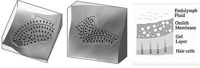
Description English: The
otoliths are the human sensory organs for linear acceleration. The utricle (left)
is approximately horizontally oriented; the saccule (center) lies approximately
vertical. The arrows indicate the local on-directions of the hair cells; and
the thick black lines indicate the location of the striola. On the right you
see a cross-section through the otolith membrane. The graphs have been
generated by Rudi Jaeger, while we cooperated on investigations of the otolith
dynamics.
Date 15 February 2011
Source Own work
Author Thomas.haslwanter
Licensing
I, the copyright holder of this work, hereby publish it
under the following licenses:
This file is
licensed under the Creative commons Attribution-Share Alike 3.0 Unported license.
You are free:
·
to share – to copy,
distribute and transmit the work
·
to remix – to adapt the work
Under the following conditions:
·
attribution – You must give
appropriate credit, provide a link to the license, and indicate if changes were
made. You may do so in any reasonable manner, but not in any way that suggests
the licensor endorses you or your use.
share alike – If you remix,
transform, or build upon the material, you must distribute your contributions
under the same or compatible license as the original.
Permission is granted to copy, distribute and/or
modify this document under the terms of the GNU Free Documentation License, Version 1.2 or any
later version published by the Free Software Foundation; with no Invariant Sections, no Front-Cover
Texts, and no Back-Cover Texts. A copy of the license is included in the
section entitled GNU Free Documentation License.
You may select the license of your choice.
Modified: A.G. DiGiovanna
Added labels and
lines. Blocked some portions.