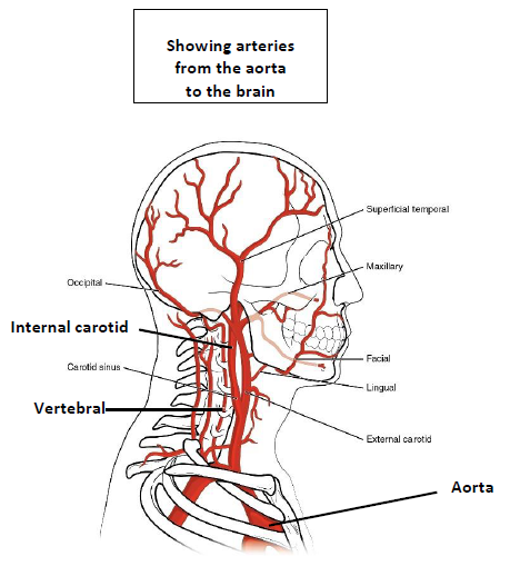
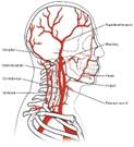
Fig. 6.9 Blood vessels that supply the brain. Scroll down. (Sources of images below. Used with permission.)
©
Copyright 2020: Augustine G. DiGiovanna, Ph.D.,
Salisbury University, Maryland
The materials on this site are licensed under CC BY-NC-SA
4.0
![]()
Attribution-NonCommercial-ShareAlike
This license requires that reusers
give credit to the creator. It allows reusers
to distribute, remix, adapt, and build upon the material in any medium
or format, for noncommercial purposes only. If others modify or adapt
the material, they must license the modified material under identical
terms.
Previous print editions of the text Human Aging: Biological Perspectives
are © Copyright 2000, 1994 by The McGraw-Hill Companies, Inc. and 2020
by Augustine DiGiovanna.
View License Deed |
View Legal Code
Sources of images. Used with permission.
https://upload.wikimedia.org/wikipedia/commons/1/12/2122_Common_Carotid_Artery.jpg
https://commons.wikimedia.org/wiki/File:2122_Common_Carotid_Artery.jpg
Description Illustration from Anatomy & Physiology, Connexions Web site. http://cnx.org/content/col11496/1.6/, Jun 19, 2013.
Source Anatomy & Physiology, Connexions Web site. http://cnx.org/content/col11496/1.6/, Jun 19, 2013.
Author OpenStax College
Licensing
This file is licensed under the Creative Commons Attribution 3.0 Unported license.
You are free:
· to share – to copy, distribute and transmit the work
· to remix – to adapt the work
Under the following conditions:
attribution – You must give appropriate credit, provide a link to the license, and indicate if changes were made. You may do so in any reasonable manner, but not in any way that suggests the licensor endorses you or your use.
Modified: A.G. DiGiovanna
Added labels
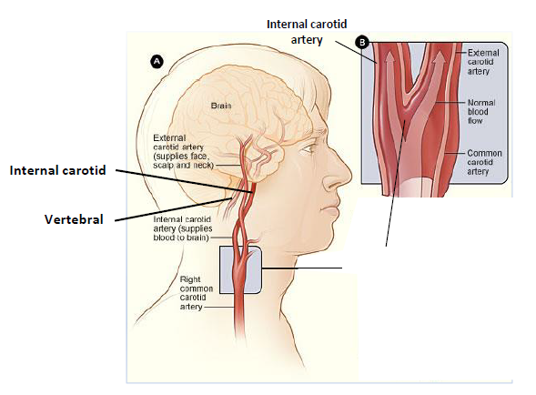
https://commons.wikimedia.org/wiki/File:Cad_anatomy.jpg
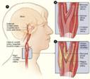
Description English: Figure A shows the location of the right carotid artery in the head and neck. Figure B is a cross-section of a normal carotid artery that has normal blood flow. Figure C shows a carotid artery that has plaque buildup and reduced blood flow.
Date 12 November 2013, 23:11:23
Source National Heart Lung and Blood Insitute (NIH)
Author National Heart Lung and Blood Insitute (NIH)
Licensing
This work is in the public domain in the United States because it is a work prepared by an officer or employee of the United States Government as part of that person’s official duties under the terms of Title 17, Chapter 1, Section 105 of the US Code. Note: This only applies to original works of the Federal Government and not to the work of any individual U.S. state, territory, commonwealth, county, municipality, or any other subdivision. This template also does not apply to postage stamp designs published by the United States Postal Service since 1978. (See § 313.6(C)(1) of Compendium of U.S. Copyright Office Practices). It also does not apply to certain US coins; see The US Mint Terms of Use.
Modified: A.G. DiGiovanna
Added labels and blocked some content
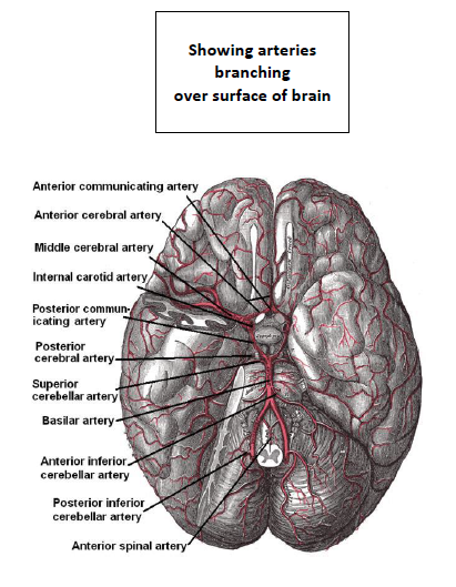
https://commons.wikimedia.org/wiki/File:Arteries_beneath_brain_Gray_closer.jpg
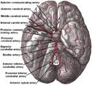
Description The brain and arteries at base of the brain. Circle of Willis is formed near center. The temporal pole of the cerebrum and a portion of the cerebellar hemisphere have been removed on the right side. Inferior aspect (viewed from below).
Date 3 February 2008
Source Derivative of Image:Gray516.png, narrowing labels to magnify image 16%; removed smudges near labels.
Author Wikid77
Other versions Derivative works of this file:
- Higher-resolution view with wider labels: Image:Arteries beneath brain.png.
Licensing
Unless stated otherwise, this image is from the 20th U.S. edition of Gray's Anatomy of the Human Body, originally published in 1918 and therefore lapsed into the public domain. A copy of Gray's Anatomy can be found on Bartleby and also on Yahoo!.
This image is in the public domain because it is a mere mechanical scan or photocopy of a public domain original, or – from the available evidence – is so similar to such a scan or photocopy that no copyright protection can be expected to arise. The original itself is in the public domain for the following reason:
This work is in the public domain in its country of origin and other countries and areas where the copyright term is the author's life plus 100 years or fewer.
This work is in the public domain in the United States because it was published (or registered with the U.S. Copyright Office) before January 1, 1926.
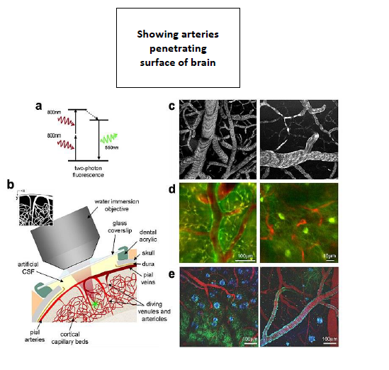
https://commons.wikimedia.org/wiki/File:Two-photon_microscopy_of_in_vivo_brain_function.jpg
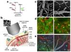
Description English: Two-photon microscopy of in vivo brain function. (a) Basic mechanism of two-photon fluorescence. (b) Schematic of surgical preparation of exposed cortex, with sealed glass window and microscope objective positioning. Green dot shows location of two-photon fluorescence. (c) Examples of two-photon maps of the vasculature following intravenous injection of dextran-conjugated fluorescein. Black dots and stripes show red blood cell motion. (d) Dual-channel imaging of neuronal (green) and vascular (red) signals: (left) Oregon Green 488 BAPTA-1 AM calcium sensitive dye stained neurons and (right) transgenic mouse expressing green fluorescent protein (GFP) in a subpopulation of neurons (mouse supplied by Jeffrey M. Friedman, Rockefeller University, New York) [101]. Texas dextran red is the intravascular tracer in both cases. (e) Three channel imaging of Tg2576 APP Alzheimer's disease mouse model with amyloid-targeting dye (blue), GFP expressing neurons and dendrites (green) and vasculature (red). Adapted from [52] and contributed by Elizabeth Hillman (Columbia University, New York).
Date 2008
Source Kherlopian et al. "A review of imaging techniques for systems biology". BMC Systems Biology 2008 2:74 doi:10.1186/1752-0509-2-74
Author Armen R Kherlopian, Ting Song, Qi Duan, Mathew A Neimark, Ming J Po, John K Gohagan and Andrew F Laine
Permission
(Reusing
this file) © 2008 Kherlopian et al;
licensee BioMed Central Ltd.
This is an Open Access article distributed under the terms of the Creative Commons Attribution License (https://creativecommons.org/licenses/by/2.0), which permits unrestricted use, distribution, and reproduction in any medium, provided the original work is properly cited.
This file is licensed under the
Creative Commons
Attribution 2.0 Generic
license.
You are free:
· to share – to copy, distribute and transmit the work
· to remix – to adapt the work
Under the following conditions:
attribution – You must give appropriate credit, provide a link to the license, and indicate if changes were made. You may do so in any reasonable manner, but not in any way that suggests the licensor endorses you or your use.
Modified: A.G. DiGiovanna
Added labels and blocked some content
_(14777725651).jpg)
Description English:
Identifier:
journaloflaborat01cent (find
matches)
Title:
The Journal of laboratory and clinical medicine
Year:
1915 (1910s)
Authors:
Central Society for Clinical Research (U.S.)
Subjects:
Biological Assay
Diagnosis, Laboratory
Disease
Medicine
Medicine
Research
Publisher:
St. Louis, C. V. Mosby
Contributing Library:
Gerstein - University of Toronto
Digitizing Sponsor:
University of Toronto
View Book Page:
Book Viewer
About This Book:
Catalog Entry
View All Images:
All Images From Book
Click here to
view book online to see this
illustration in context in a browseable online version of this book.
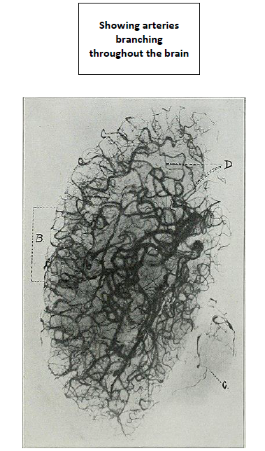
Text Appearing Before Image:
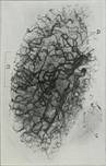
presenting both a normal and an abnormalspecimen for differential study. To
illustrate the results of this method, photographs of two plates are pre-sented
(Figs 2 and 3). There was a clinical diagnosis of cerebral hemorrhage. The
autopsy diag-nosis made without sectioning the brain was arteriosclerosis;
dififuse subarach-noid hemorrhage from the right and left posterior cerebellar
arteries; throm- Radiographic Studies of Cerebral Vascular Lesions 839 bosis of
the right and left posterior cerebellar arteries. After the exposure tothe x-ray
the brain was sectioned, showing, a mass of clotted blood which en-tirely filled
the left lateral ventricle; an area of white softening in the lefthemisphere
corresponding to that portion of the medulla which is supplied byterminal
branches of the ascending frontal artery. There were no pathologicchanges in the
right cerebrum. The x-ray diagnosis was arteriosclerosis, thevessels showing
marked tortuosity and irregularity; rupture of the left middle
Text Appearing After Image:
Fig. 3-A.—Case No. A-2997. cerebral artery at a point in its m;iin trunk, as
shown by an apparent projectioninto the intima of this artery of a part of the
clot, when scon stereoscopically;thrombosis of the posterior cerebellar
arteries, as seen by the absence of the in-jection mass in these vessels;
thrombosis of the terminal liranchcs of the ascendingfrontal artery on the left
side, as seen by the absence oi iho injection mass inthese vessels; possible
multiple miliary aneurisnial formations in the rightcerelirum. To demonstrate
these findings. I ha\ e marked the print as follows: 840 The Journal of
Laboratory and Clinical Medicine (A) Area of thrombosis of the terminal branches
of the ascending frontalbranch of the left middle cerebral; (B) Correspondinjr
normal area in the rightside; (C) Thrombosis of the right and left posterior
cerebellar arteries; (D)Possible miliary aneurismal formations.
Note About Images
Please note that these images are extracted from scanned page images that may have been digitally enhanced for readability - coloration and appearance of these illustrations may not perfectly resemble the original work.
Date 1915
Source https://www.flickr.com/photos/internetarchivebookimages/14777725651/
· Source book page: https://archive.org/stream/journaloflaborat01cent/journaloflaborat01cent#page/n848/mode/1up
Author Internet Archive Book Images
Permission
(Reusing
this file)
At
the time of upload, the image license was automatically confirmed using the
Flickr
API.
For more information see
Flickr API detail.
- Flickr tags bookid:journaloflaborat01cent
- bookyear:1915
- bookdecade:1910
- bookcentury:1900
- booksubject:Biological_Assay
- booksubject:Diagnosis__Laboratory
- booksubject:Disease
- booksubject:Medicine
- booksubject:Research
- bookpublisher:St__Louis__C__V__Mosby
- bookcontributor:Gerstein___University_of_Toronto
- booksponsor:University_of_Toronto
- bookleafnumber:848
- bookcollection:gerstein
- bookcollection:toronto
· bookcollection:medicalheritagelibrary
Flickr posted date 30 July 2014
The categories of this image need checking. You can do so here.
_(14777725651).jpg&action=edit&withJS=MediaWiki:Catcheck.js)
- Please remove redundant categories and try to put this image in the most specific category/categories.
· You can remove this template by clicking here (or on the first line).
Licensing
This image was taken from Flickr's The Commons. The uploading organization may have various reasons for determining that no known copyright restrictions exist, such as:
- The copyright is in the public domain because it has expired;
- The copyright was injected into the public domain for other reasons, such as failure to adhere to required formalities or conditions;
- The institution owns the copyright but is not interested in exercising control; or
- The institution has legal rights sufficient to authorize others to use the work without restrictions.
More information can be found at https://flickr.com/commons/usage/.
Please add additional copyright tags to this image if more specific information about copyright status can be determined. See Commons:Licensing for more information.
This image was originally posted to Flickr by Internet Archive Book Images at https://flickr.com/photos/126377022@N07/14777725651. It was reviewed on 2 November 2015 by FlickreviewR and was confirmed to be licensed under the terms of the No known copyright restrictions.
Modified: A.G. DiGiovanna
Added labels