%20motor%20pathway%20-%2007-10-21%20-%20b-snp2.PNG)
©
Copyright 2020: Augustine G. DiGiovanna, Ph.D.,
Salisbury University, Maryland
The materials on this site are licensed under CC BY-NC-SA
4.0
![]()
Attribution-NonCommercial-ShareAlike
This license requires that reusers
give credit to the creator. It allows reusers
to distribute, remix, adapt, and build upon the material in any medium
or format, for noncommercial purposes only. If others modify or adapt
the material, they must license the modified material under identical
terms.
Previous print editions of the text Human Aging: Biological Perspectives
are © Copyright 2000, 1994 by The McGraw-Hill Companies, Inc. and 2020
by Augustine DiGiovanna.
View License Deed |
View Legal Code
Sources of
images. Used with permission.
%20motor%20pathway%20-%2007-10-21%20-%20b-snp_files/image002.gif)
https://commons.wikimedia.org/wiki/File:Motor_homunculus.svg
Description English: The "motor homunculus". The
body surface is projected onto the gyrus precentralis
(coronal section of the right hemisphere).
Date 11
June 2016
Source
File:Homunculus-ja.png
After Penfield and Rasmussen (1950), The Cerebral Cortex of Man.
Modified
from (an earlier version of) File:Homunculus-de.png by Was a bee.
Author
mailto:ralf@ark.in-berlin.de
Licensing
This file is
licensed under the
Creative Commons
Attribution-Share Alike 4.0 International license.
You are free:
·
to share – to copy, distribute and transmit the work
·
to remix – to adapt the work
Under the following conditions:
·
attribution – You must give appropriate credit, provide a link to the license, and
indicate if changes were made. You may do so in any reasonable manner, but not
in any way that suggests the licensor endorses you or your use.
share alike – If you remix, transform,
or build upon the material, you must distribute your contributions under the same or compatible license as the original.
%20motor%20pathway%20-%2007-10-21%20-%20b-snp_files/image004.jpg)
https://upload.wikimedia.org/wikipedia/commons/7/78/Precentral_gyrus_-_superior_view2.png
https://commons.wikimedia.org/wiki/File:Precentral_gyrus_-_superior_view2.png
Description English: Precentral gyrus (shown in red).
Date 17
December 2012
Source
en:Anatomography (setting page of this image)
Author
Anatomography
This file is licensed under
the Creative Commons Attribution-Share Alike 2.1
Japan license.
You are free:
·
to share – to copy, distribute and transmit the work
·
to remix – to adapt the work
Under the following conditions:
·
attribution – You must give appropriate credit, provide a link to the license, and
indicate if changes were made. You may do so in any reasonable manner, but not
in any way that suggests the licensor endorses you or your use.
share alike – If you remix, transform,
or build upon the material, you must distribute your contributions under the same or compatible license as the original.
This image was made out of,
or made from, content published in a BodyParts3D/Anatomography web site. The content of their website is published under the Creative Commons Attribution 2.1 Japan license. The
author and licenser of the contents is
- "BodyParts3D, ©
The Database Center for Life Science licensed under CC Attribution-Share
Alike 2.1 Japan." (Google translate)
You can download 3D-polygon data of whole human body. And
you can also manipulate and edit the polygon data using 3D softwares,
for example,
Meshlab or
Blender.
%20motor%20pathway%20-%2007-10-21%20-%20b-snp_files/image006.jpg)
https://en.wikipedia.org/wiki/File:Gray704.png
https://en.wikipedia.org/wiki/Cerebellar_tonsil
Description This file has no description,
and may be lacking other information.
Please provide a meaningful
description of this file.
Plate 704
Date before
1858
- Source
Henry Gray (1918) Anatomy of the Human Body
(See "Book" section below)
·
Bartleby.com: Gray's Anatomy, Plate 704
Author
Henry Vandyke Carter (1831–1897)
Book
Henry Gray: Gray's Anatomy (20th edition)
Author
Henry Gray
(1827–1861)
-_Title_page.png)
Editor
Revised by Warren H. Lewis
Illustrator Henry Vandyke Carter (1831–1897)
Title
Anatomy of the Human Body
Edition
20
Publisher
Lea and Febiger
Object type version, edition,
or translation
Page overview
list of all the
plates
Language English
Publication date
1918
Place of publication Philadelphia and New York
Source
Bartleby
Licensing
This image is in the
public domain because it is a mere
mechanical scan or photocopy of a public domain original, or – from the
available evidence – is so similar to such a scan or photocopy that no
copyright protection can be expected to arise. The original itself is in the
public domain for the following reason:
This work is in the
public domain in its country of origin and other countries and areas where the
copyright term is the author's life
plus 100 years or fewer.
This work is in
the
public domain in the
United States because it was
published (or registered with the
U.S. Copyright Office) before January 1, 1926.
%20motor%20pathway%20-%2007-10-21%20-%20b-snp_files/image008.jpg)
https://upload.wikimedia.org/wikipedia/commons/9/96/Biceps_Muscle_CNX.jpg
https://commons.wikimedia.org/wiki/File:Biceps_Muscle_CNX.jpg
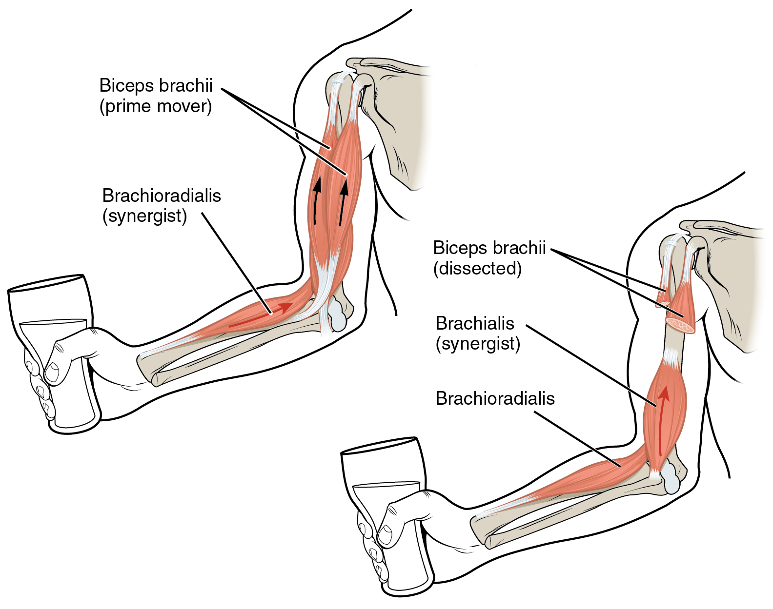
Description English: The biceps brachii flex the lower arm.
The brachoradialis, in the forearm, and brachialis,
located deep to the biceps in the upper arm, are both synergists that aid in
this motion.
Date 23
May 2013, 20:16:05
Source
http://cnx.org/content/m46487/latest/?collection=col11496/latest
Author
OpenStax College
Licensing
This file is
licensed under the
Creative Commons
Attribution-Share Alike 3.0
Unported license.
You are free:
·
to share – to copy, distribute and transmit the work
·
to remix – to adapt the work
Under the following conditions:
·
attribution – You must give appropriate credit, provide a link to the license, and
indicate if changes were made. You may do so in any reasonable manner, but not
in any way that suggests the licensor endorses you or your use.
share alike – If you remix, transform,
or build upon the material, you must distribute your contributions under the same or compatible license as the original.
I, the copyright holder of this work, hereby publish it under the following
license:
This
file is licensed under the Creative Commons Attribution 4.0
International license.
You are free:
·
to share – to copy, distribute and transmit the work
·
to remix – to adapt the work
Under the following conditions:
attribution – You must give appropriate
credit, provide a link to the license, and indicate if changes were made. You
may do so in any reasonable manner, but not in any way that suggests the
licensor endorses you or your use.