Fig. 6.11 Alzheimer’s disease (Sources of images and videos below. Used with permission.)


“Alzheimer disease”
https://medlineplus.gov/ency/anatomyvideos/000003.htm
“Alzheimers Disease”
https://blausen.com/en/video/alzheimers-disease/
“Video: How Alzheimer's Changes the Brain”
©
Copyright 2020: Augustine G. DiGiovanna, Ph.D.,
Salisbury University, Maryland
The materials on this site are licensed under CC BY-NC-SA
4.0
![]()
Attribution-NonCommercial-ShareAlike
This license requires that reusers
give credit to the creator. It allows reusers
to distribute, remix, adapt, and build upon the material in any medium
or format, for noncommercial purposes only. If others modify or adapt
the material, they must license the modified material under identical
terms.
Previous print editions of the text Human Aging: Biological Perspectives
are © Copyright 2000, 1994 by The McGraw-Hill Companies, Inc. and 2020
by Augustine DiGiovanna.
View License Deed |
View Legal Code
Sources of
images and videos. Used with permission.

https://live.staticflickr.com/1594/24524716351_f762c70a64_b.jpg
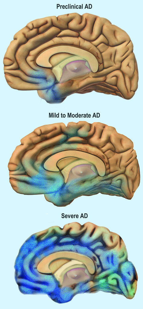
Description English: Alzheimers disease progression.
Date 8
March 2013, 14:20:38
Source http://www.nia.nih.gov/alzheimers/topics/alzheimers-basics
Author National
Institute on Aging
Licensing
This
work is in the public domain in the United States because
it is a work prepared by
an officer or employee of the United States Government as part of that person’s
official duties under the terms of Title
17, Chapter 1, Section 105 of the US Code. Note: This only applies to original works of the Federal
Government and not to the work of any individual U.S.
state, territory, commonwealth, county,
municipality, or any other subdivision. This template also does not apply to
postage stamp designs published by the United States Postal Service since 1978. (See § 313.6(C)(1)
of Compendium of U.S. Copyright Office Practices). It also does not apply to
certain US coins; see The
US Mint Terms of Use.

https://live.staticflickr.com/1466/24239522109_80aa4defd1_z.jpg
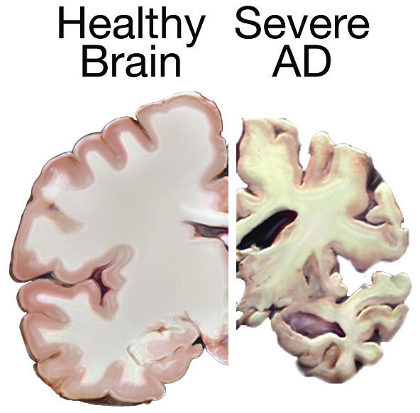
Description English: A healthy brain compared to a
brain suffering from Alzheimer's Disease
Deutsch: Gesundes Gehirn (links) im Vergleich mit dem Gehirn eines
Menschen mit Alzheimer-Demenz (rechts)
Date 13
October 2011
Source https://www.nlm.nih.gov/medlineplus/magazine/issues/fall10/articles/fall10pg20-21.html
Author National
Institutes of Health
Licensing
This image is a work of the National Institutes of Health,
part of the United
States Department of Health and Human Services. As a work of the U.S. federal government,
the image is in the public domain.

https://commons.wikimedia.org/wiki/File:Alzheimer%27s_disease_brain_comparison.jpg
Description Combination of two brain diagrams
in one for comparison. In the left normal brain, in the right brain of a person
with Alzheimer's disease
English: Diagram of the brain of a person with Alzheimer's Disease
English: Diagram of a normal brain
·
Source SEVERESLICE_HIGH.JPG
·
Author derivative
work: Garrondo (talk)
·
SEVERESLICE_HIGH.JPG: ADEAR:
"Alzheimer's Disease Education and Referral Center, a service of the
National Institute on Aging."
·
PRECLINICALSLICE_HIGH.JPG: ADEAR:
"Alzheimer's Disease Education and Referral Center, a service of the
National Institute on Aging."
Other versions Derivative works of this file:
·
Cerebro corte sagital
Alzheimer.jpg
·
Alzheimer's disease
brain comparison-ar.jpg
·
Alzheimer's disease
brain comparison-zh.jpg
This is a retouched picture, which means that it has
been digitally altered from its original version. Modifications: Combined
two images in one. The original can be viewed here: PRECLINICALSLICE HIGH.JPG: ![]() . Modifications made by Garrondo.
. Modifications made by Garrondo.
I, the copyright holder of this work, release this work
into the public domain. This applies worldwide.
In some countries
this may not be legally possible; if so:
I grant anyone the right to use this work for any purpose, without
any conditions, unless such conditions are required by law.
Original upload log
This image is a derivative work of the following images:
Mark in bkini
·
Image:SEVERESLICE_HIGH.JPG licensed with
PD-USGov
o
2008-07-30T13:23:22Z Garrondo
3000x2250 (4045486 Bytes) {{Information |Description={{en|1=Diagram of the
brain of a person with Alzheimer's Disease}}
|Source=http://www.nia.nih.gov/Alzheimers/Resources/MediaRoom.htm
|Author=ADEAR: "Alzheimer's Disease Education and Referral Cente
·
Image:PRECLINICALSLICE_HIGH.JPG
licensed with PD-USGov
o
2008-07-30T13:15:53Z Garrondo
2778x2208 (4096555 Bytes) {{Information |Description={{en|1=Diagram of a
normal brain}}
|Source=http://www.nia.nih.gov/Alzheimers/Resources/MediaRoom.htm |Author=ADEAR:
"Alzheimer's Disease Education and Referral Center, a service of the
National Ins
uploaded with derivativeFX
Modified: A.G.DiGiovanna
Blocked portions of image

https://commons.wikimedia.org/wiki/File:Hippocampus_image.png
Description English: hippocampus. Images are from
Anatomography maintained by Life Science Databases(LSDB).
Life Science Databases(LSDB)のAnatomography
Date 25
September 2009
Source from Anatomography, website maintained
by Life Science Databases(LSDB).
You can get this image through URL below.
URL.
Author Images
are generated by Life Science Databases(LSDB).
Permission
(Reusing this file) CC-BY-SA-2.1-jp
This file is licensed under the Creative commons Attribution-Share
Alike 2.1 Japan license.
You are free:
·
to share – to copy,
distribute and transmit the work
·
to remix – to adapt
the work
Under the following conditions:
·
attribution – You must
give appropriate credit, provide a link to the license, and indicate if changes
were made. You may do so in any reasonable manner, but not in any way that
suggests the licensor endorses you or your use.
share alike – If you remix, transform, or build upon the material,
you must distribute your contributions under the same
or compatible license as the original.
This image was made out of, or made from, content
published in a BodyParts3D/Anatomography web site. The
content of their website
is published under the Creative commons Attribution 2.1 Japan license. The
author and licenser of the contents is
·
"BodyParts3D,
© The Database Center for Life Science licensed under CC Attribution-Share
Alike 2.1 Japan." (Google
translate)
You can
download 3D-polygon data of whole human body. And you can also manipulate and
edit the polygon data using 3D softwares, for example, Meshlab or Blender.
·
Download
BodyParts3D Human Body Polygon Data
Modified: A.G.DiGiovanna
Added labels and pointers

https://commons.wikimedia.org/wiki/File:Hippocampus_small.gif
Description English: hippocampus. Images are from
Anatomography maintained by Life Science Databases(LSDB).
Life Science Databases(LSDB)のAnatomography
Date 23
March 2010
Source from Anatomography, website maintained
by Life Science Databases(LSDB).
You can get this image through URL below.
URL.
Author Images
are generated by Life Science Databases(LSDB).
Permission
(Reusing this file) CC-BY-SA-2.1-jp
Other versions

larger one This file is licensed under the Creative commons Attribution-Share
Alike 2.1 Japan license.
You are free:
·
to share – to copy,
distribute and transmit the work
·
to remix – to adapt
the work
Under the following conditions:
·
attribution – You must
give appropriate credit, provide a link to the license, and indicate if changes
were made. You may do so in any reasonable manner, but not in any way that
suggests the licensor endorses you or your use.
share alike – If you remix, transform, or build upon the material,
you must distribute your contributions under the same
or compatible license as the original.
This image was
made out of, or made from, content published in a BodyParts3D/Anatomography
web site. The content of their website
is published under the Creative commons Attribution 2.1 Japan license. The
author and licenser of the contents is
-
"BodyParts3D,
© The Database Center for Life Science licensed under CC Attribution-Share
Alike 2.1 Japan." (Google
translate)
You can download 3D-polygon data
of whole human body. And you can also manipulate and edit the polygon data
using 3D softwares, for example, Meshlab or Blender.
·
Download
BodyParts3D Human Body Polygon Data

https://live.staticflickr.com/4557/38686503251_f0bf300990_b.jpg
https://www.flickr.com/photos/nihgov/38686503251
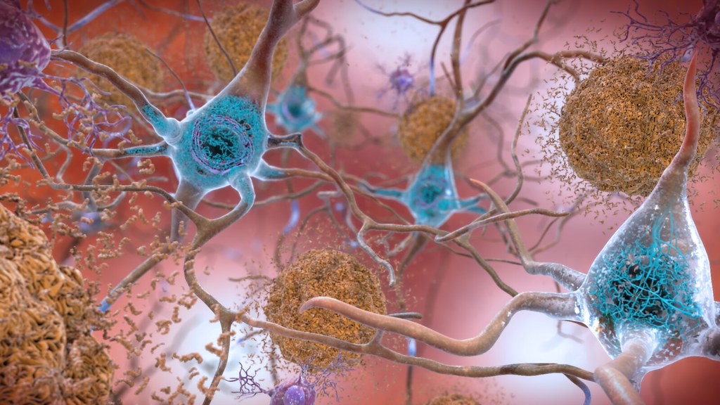
“In the Alzheimer’s
affected brain, abnormal levels of the beta-amyloid protein clump together to
form plaques (seen in brown) that collect between neurons and disrupt cell
function. Abnormal collections of the tau protein accumulate and form tangles
(seen in blue) within neurons, harming synaptic communication between nerve
cells.”
Beta-Amyloid
Plaques and Tau in the Brain
https://www.flickr.com/photos/nihgov/25209610730/in/album-72157662951050375/
VIDEOS
“Alzheimer disease”
https://medlineplus.gov/ency/anatomyvideos/000003.htm

https://live.staticflickr.com/4557/38686503251_f0bf300990_b.jpg
“In the Alzheimer’s affected brain, abnormal levels of
the beta-amyloid protein clump together to form plaques (seen in brown) that
collect between neurons and disrupt cell function. Abnormal collections of the
tau protein accumulate and form tangles (seen in blue) within neurons, harming
synaptic communication between nerve cells.”
Beta-Amyloid
Plaques and Tau in the Brain
https://www.flickr.com/photos/nihgov/38686503251
“In the Alzheimer’s affected brain, abnormal levels of
the beta-amyloid protein clump together to form plaques (seen in brown) that
collect between neurons and disrupt cell function. Abnormal collections of the
tau protein accumulate and form tangles (seen in blue) within neurons, harming
synaptic communication between nerve cells.”
Beta-Amyloid
Plaques and Tau in the Brain
https://www.flickr.com/photos/nihgov/25209610730/in/album-72157662951050375/

https://upload.wikimedia.org/wikipedia/commons/9/99/Alzheimers_Disease.jpg
VIDEOS
“Alzheimer
disease”
https://medlineplus.gov/ency/anatomyvideos/000003.htm
“Alzheimers
Disease”
https://blausen.com/en/video/alzheimers-disease/
“Video:
How Alzheimer's Changes the Brain”