Fig. 6.10 Strokes. Scroll down. (Sources of images and videos below. Used
with permission.)
First view of ischemic stroke from emboli
Second view of ischemic stroke from emboli
First view of hemorrhagic stroke from aneurysms
Second view of hemorrhagic stroke from aneurysms
One view of diffusing hemorrhagic stroke
Videos
Stroke
https://blausen.com/en/video/stroke/
Abnormal
Hemostasis: Embolus Causes a Stroke
https://blausen.com/en/video/abnormal-hemostasis-embolus-causes-a-stroke/
Sources of images and videos. Used
with permission.
Stroke
https://blausen.com/en/video/stroke/
Abnormal
Hemostasis: Embolus Causes a Stroke
https://blausen.com/en/video/abnormal-hemostasis-embolus-causes-a-stroke/
©
Copyright 2020: Augustine G. DiGiovanna, Ph.D.,
Salisbury University, Maryland
The materials on this site are licensed under CC BY-NC-SA
4.0
![]()
Attribution-NonCommercial-ShareAlike
This license requires that reusers
give credit to the creator. It allows reusers
to distribute, remix, adapt, and build upon the material in any medium
or format, for noncommercial purposes only. If others modify or adapt
the material, they must license the modified material under identical
terms.
Previous print editions of the text Human Aging: Biological Perspectives
are © Copyright 2000, 1994 by The McGraw-Hill Companies, Inc. and 2020
by Augustine DiGiovanna.
View License Deed |
View Legal Code
Sources of images and videos. Used with permission.
https://commons.wikimedia.org/wiki/File:Ischemic_Stroke.svg
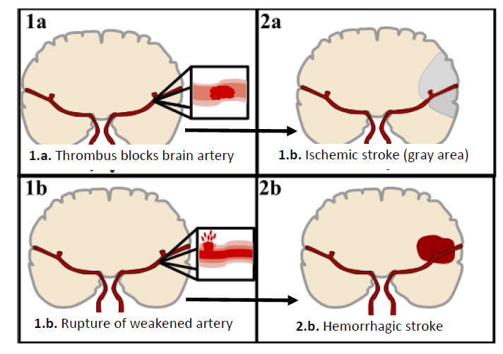
Description English: An ischemic stroke results from a
clot forming in one of the brain’s arteries blocking the blood flow to a
specific portion of the brain. This results in the death of the affected brain
cells if the clot isn’t dealt with in time.
1. This is a
cross section of the brain allowing for the main arteries supplying blood to
the brain to be seen.
2. The artery
of the brain which carries nutrient containing blood from the heart and lungs
to the brain thus allowing for energy creation by the brain cells to keep them
alive.
3. The clot, a
concentration of blood cells in the artery, blocks the flow of blood to the
brain making it hard for the nutrients to reach the brain cells, and after many
minutes, the eventual death of the brain cells.
4. Tributary
arteries supply blood to the brain both under regular conditions and in the
occurrence of a clot in the main blood supplying arteries.
The result of an ischemic stroke is individualized
depending on where in the brain the clot formed and how severe the brain damage
was. Common disabilities as a result of an ischemic stroke are speech and
vision disabilities, physical disabilities, memory loss, and behavior changes.
Date 3
June 2018
Source Own work
Author Elinor Hunt
Licensing
I, the copyright holder of this work,
hereby publish it under the following license:
This file is licensed under the Creative commons Attribution-Share
Alike 4.0 International license.
You are free:
·
to
share – to copy,
distribute and transmit the work
·
to
remix – to adapt the work
Under the following conditions:
·
attribution – You must give appropriate credit,
provide a link to the license, and indicate if changes were made. You may do so
in any reasonable manner, but not in any way that suggests the licensor
endorses you or your use.
share alike – If you remix, transform, or build upon the
material, you must distribute your contributions under the same
or compatible license as the original.
Modified: A.G.DiGiovanna
Added labels and arrows
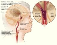
https://commons.wikimedia.org/wiki/File:Stroke_ischemic.jpg
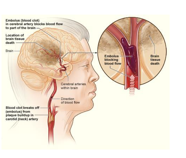
Description English: The illustration shows how an
ischemic stroke can occur in the brain. If a blood clot breaks away from plaque
buildup in a carotid (neck) artery, it can travel to and lodge in an artery in
the brain. The clot can block blood flow to part of the brain, causing brain
tissue death.
Date 12
November 2013, 21:15:53
Source National Heart Lung and Blood Institute (NIH)
Author National Heart Lung and Blood Institute (NIH)
Licensing
This
work is in the public domain in the United States because
it is a work prepared by
an officer or employee of the United States Government as part of that person’s
official duties under the terms of Title
17, Chapter 1, Section 105 of the US Code.
Note: This only applies to
original works of the Federal Government and not to the work of any individual U.S.
state, territory, commonwealth, county,
municipality, or any other subdivision. This template also does not apply to
postage stamp designs published by the United States Postal Service since 1978. (See § 313.6(C)(1)
of Compendium of U.S. Copyright Office Practices). It also does not apply to
certain US coins; see The
US Mint Terms of Use.
File history
Click on a date/time to view the file as it appeared at
that time.
Date/Time
Thumbnail Dimensions User Comment
current 20:35, 12 November 2013 475 × 381 (81 KB) CFCF (talk | contribs) User created page with upload Wizard
Modified: A.G.DiGiovanna
Added labels
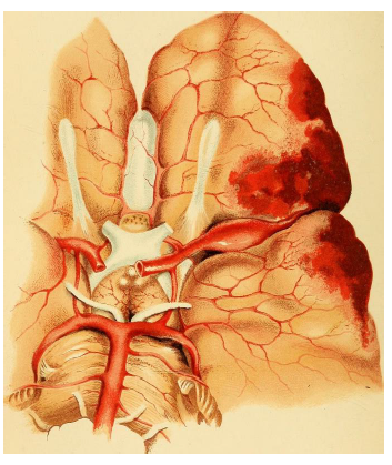
_(1870)_(14763398095).jpg)
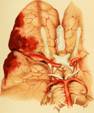
Description English:
Aneurysm of left middle cerebral artery
Identifier: onepilepsyanatom00eche (find
matches)
Title: On
epilepsy : anatomo-pathological and clinical notes (with original plates
and engravings.)
Year: 1870
(1870s)
Authors: Echeverria,
M. Gonzalez (Manuel Gonzalez)
Subjects: Epilepsy
Epilepsy
Publisher: New
York : W. Wood
Contributing Library: Francis
A. Countway Library of Medicine
Digitizing Sponsor: Open
Knowledge commons and Harvard Medical School
View Book Page: Book
Viewer
About This Book: Catalog
Entry
View All Images: All
Images From Book
Click here to view
book online to see this illustration in context in a browseable
online version of this book.
Text Appearing Before Image:
the ptosis and paralysis of the left limbs. The antisyphilitic treatment
with the iodide of potassium, proto-iodideof mercury, and cod liver oil, proved
of no more avail than to remove the cutaneous accidents. The epileptic attacks
continuing with troublesome coughing, vomiting, and other nervous symptoms, the
intellectual faculties gradually failed, and, running into a soporous condition,
the patient finally died in a fit, the 12th March, 1863.In the commencement, an
aura starting from the fingers on the left hand, preceded the paroxysms, but
subsequently this warning ceased. During the fits the girl used to bite the
tongue, and to froth considerably at the mouth. Autopsy.—Calvarium normal,
vessels of the diploe very congested, meninges opaque. Serous effusion in the
arachnoid cavity: this membrane opalescent on the anterior part of the base of
the brain. Right middle cerebral artery plugged by a firm, laminated,
orange-tinted clot, adhering to the walls and causing aneurismal dilatation
PMeV
Text Appearing After Image:
Ad riot, eop!^ Aneurism of the left middle cerebral artery. Chi-.Fr Mifller.
CkromoUih:^ Carlsruk&. OF EPILEPSY. 73 of the vessel. A large surface of
the anterior and middle lobes in the vicinity covered by a film of coagulated
blood, and the gray matter underneath, infiltrated and softened, without
delimited boundaries with the white substance somewhat yellowish in this
region. On section the brain tissue looks rather wet, with two yellow patches the
size of a hazel nut, in the right centrum ovale, near the corpus striatum, and
surrounded by softened tissue. Ventricles containing turbid serosity ;
choroid plexus rough, covered by small, firm, yellow granulations. These
miliary bodies covered also the ependyma of the fourth ventricle, and the
pia-mater over the medulla. Both lungs with tuberculous deposits at the apex,
extensive pleuritic adhesions on right side, with gray hepatization of the
lower lobe. Heart natural, without any valvular lesion. The other viscera presented
nothing worthy of
Note About Images
Please note that these images are extracted from scanned
page images that may have been digitally enhanced for readability - coloration
and appearance of these illustrations may not perfectly resemble the original
work.
Date 1870
Source https://www.flickr.com/photos/internetarchivebookimages/14763398095/
·
Source book page: https://archive.org/stream/onepilepsyanatom00eche/onepilepsyanatom00eche#page/n88/mode/1up
Author
Internet Archive Book Images
Permission
(Reusing this file)
At
the time of upload, the image license was automatically confirmed using the
Flickr API. For
more information see Flickr API detail.
·
Flickr tags bookid:onepilepsyanatom00eche
·
bookyear:1870
·
bookdecade:1870
·
bookcentury:1800
·
bookauthor:Echeverria__M__Gonzalez__Manuel_Gonzalez_
·
booksubject:Epilepsy
·
bookpublisher:New_York___W__Wood
·
bookcontributor:Francis_A__Countway_Library_of_Medicine
·
booksponsor:Open_Knowledge_commons_and_Harvard_Medical_School
·
bookleafnumber:88
·
bookcollection:medicalheritagelibrary
·
bookcollection:francisacountwaylibrary
·
bookcollection:americana
Flickr
posted date 28 July 2014
Licensing
This image was taken from Flickr's The commons. The uploading organization
may have various reasons for determining that no known copyright
restrictions exist, such as:
1.
The copyright is in the public domain because it has expired;
2.
The copyright was injected into the public domain for other reasons, such
as failure to adhere to required formalities or conditions;
3.
The institution owns the copyright but is not interested in exercising
control; or
4.
The institution has legal rights sufficient to authorize others to use the
work without restrictions.
More
information can be found at https://flickr.com/commons/usage/.
Please add additional copyright tags to this
image if more specific information about copyright status can be determined.
See commons:Licensing
for more information.
Modified: A.G.DiGiovanna
Reversed image
https://commons.wikimedia.org/wiki/File:Atrial_fib_stroke.jpg
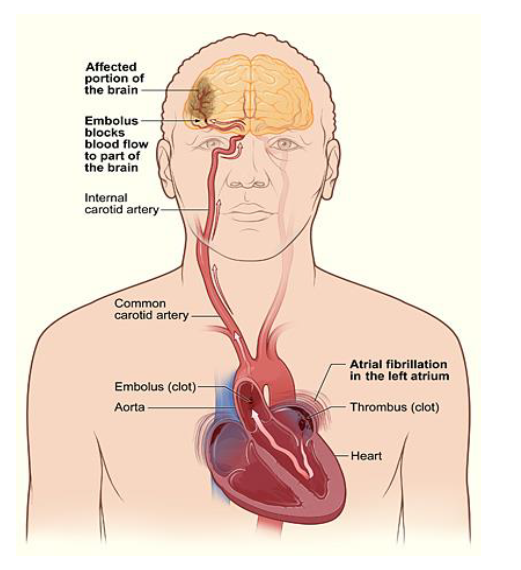
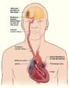
Description English: The illustration shows how a
stroke can occur during atrial fibrillation. A blood clot (thrombus) can form
in the left atrium of the heart. If a piece of the clot breaks off and travels
to an artery in the brain, it can block blood flow through the artery. The lack
of blood flow to the portion of the brain fed by the artery causes a stroke.
Date 12
November 2013, 22:58:27
Source
National
Heart Lung and Blood Institute (NIH)
Author
National
Heart Lung and Blood Institute (NIH)
Licensing
This
work is in the public domain in the United States because
it is a work prepared by
an officer or employee of the United States Government as part of that person’s
official duties under the terms of Title
17, Chapter 1, Section 105 of the US Code.
Note: This only applies to original works of the Federal
Government and not to the work of any individual U.S.
state, territory, commonwealth, county,
municipality, or any other subdivision. This template also does not apply to
postage stamp designs published by the United States Postal Service since 1978. (See § 313.6(C)(1)
of Compendium of U.S. Copyright Office Practices). It also does not apply to
certain US coins; see The
US Mint Terms of Use.
Modified: A.G.DiGiovanna
Added labels
https://commons.wikimedia.org/wiki/File:Stroke_hemorrhagic.jpg
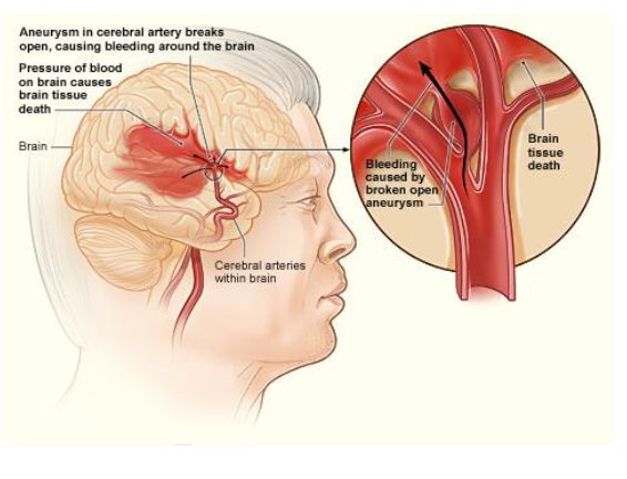
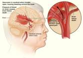
Description English: The illustration shows how a
hemorrhagic stroke can occur in the brain. An aneurysm in a cerebral artery
breaks open, which causes bleeding in the brain. The pressure of the blood
causes brain tissue death.
Date 12
November 2013, 21:15:49
Source
National
Heart Lung and Blood Insitute (NIH)
Author
National
Heart Lung and Blood Insitute (NIH)
Licensing
This
work is in the public domain in the United States because
it is a work prepared by
an officer or employee of the United States Government as part of that person’s
official duties under the terms of Title
17, Chapter 1, Section 105 of the US Code.
Note: This only applies to original works of the Federal
Government and not to the work of any individual U.S.
state, territory, commonwealth, county,
municipality, or any other subdivision. This template also does not apply to
postage stamp designs published by the United States Postal Service since 1978. (See § 313.6(C)(1)
of Compendium of U.S. Copyright Office Practices). It also does not apply to
certain US coins; see The
US Mint Terms of Use.
Modified: A.G.DiGiovanna
Added labels
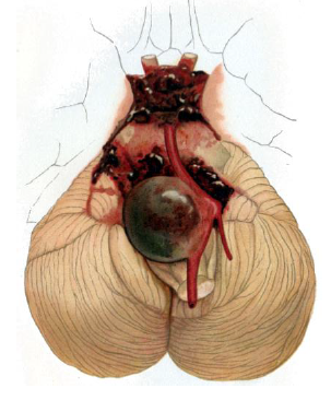
https://commons.wikimedia.org/wiki/File:Cerebellar_aneurysm.png
Description
Deutsch:
Aneurysma der Arteria basilaris und der Arteriae
vertebralis
English:
Aneurysm of the basilar artery and the vertebral arteries
Date 10
December 2013, 16:21:57
Source LEHMANN'S MEDIC IN. HAND ATLANTA Atlas
und Grundriss der PATHOLOGIC
ANATOMIZE
1901
Author
Professor
Dr. O. Bollinger.
Licensing
This
is a faithful photographic reproduction of a two-dimensional, public
domain work of art. The work of art itself is in the public domain for the
following reason:
This work is in the public
domain in its country of origin and other countries and areas where the
copyright term is the
author's life plus 70 years or fewer.
You must also include a United States public
domain tag to indicate why this work is in the public domain in the United
States. Note that a few countries have copyright terms longer than 70
years: Mexico has 100 years, Jamaica has 95 years, Colombia has 80 years, and
Guatemala and Samoa have 75 years. This image may not be in the public
domain in these countries, which moreover do not implement the rule of the shorter term. Côte d'Ivoire
has a general copyright term of 99 years and Honduras has 75 years, but they do
implement the rule of the shorter term. Copyright may extend on works created
by French who died for France in World
War II (more information),
Russians who served in the Eastern Front of World War II
(known as the Great Patriotic War in Russia) and posthumously rehabilitated victims of Soviet
repressions (more information).
Modified: A.G.DiGiovanna
Added labels