Fig. 2.5 The hierarchy of body structure (Images used with permission. See below and
separate page)
Copyright
2020: Augustine G. DiGiovanna, Ph.D., Salisbury University, Maryland
The materials on this site are licensed under CC BY-NC-SA 4.0 .

https://www.biologyofhumanaging.com/Figures/CC-BY-NS-SA%20image.jpg
Attribution-NonCommercial-ShareAlike
This license requires that reusers
give credit to the creator. It allows reusers to
distribute, remix, adapt, and build upon the material in any medium or format,
for noncommercial purposes only. If others modify or adapt the material, they
must license the modified material under identical terms.
Previous print editions of the text Human Aging: Biological Perspectives are ©
Copyright 2000, 1994 by The McGraw-Hill Companies, Inc. and 2020 by Augustine
DiGiovanna.
View License Deed | View Legal Code
Sources of images used.
Atoms
https://commons.wikimedia.org/wiki/File:Atom_clipart_violet.svg
https://upload.wikimedia.org/wikipedia/commons/thumb/8/86/Atom_clipart_violet.svg/184px-Atom_clipart_violet.svg.png
Description Description:
Atom Structure of Beryllium (e=4, P=4, n=4) in violet Theme. Source: Own work
Date 9 August 2010, 22:49 (UTC)
Source Atom_clipart_violet.png
Author Atom_clipart_violet.png: Teetaweepo
derivative work: Marek M (talk)
Molecules
https://upload.wikimedia.org/wikipedia/commons/thumb/5/5f/Water_molecule_3D_with-signs.svg/600px-Water_molecule_3D_with-signs.svg.png
https://commons.wikimedia.org/wiki/File:Water_molecule_3D_with-signs.svg
https://upload.wikimedia.org/wikipedia/commons/thumb/5/5f/Water_molecule_3D_with-signs.svg/600px-Water_molecule_3D_with-signs.svg.png
Description English:
Molecule of water.
Date 3 March 2020, 21:59 (UTC)
Source This file was derived
from: Water
molecule 3D.svg:
Author File:Water
molecule 3D.svg:
derivative work: MikeRun
https://upload.wikimedia.org/wikipedia/commons/thumb/1/1c/Water_molecule_3D.svg/600px-Water_molecule_3D.svg.png
https://commons.wikimedia.org/wiki/File:Water_molecule_3D.svg
Description English: Molecule
of water.
Date 2
November 2006
Source Water-3D-vdW.png
Author Dbc334 (first version); Jynto (second version)
https://commons.wikimedia.org/wiki/File:DNA-fragment-3D-vdW.png
https://upload.wikimedia.org/wikipedia/commons/thumb/2/2c/DNA-fragment-3D-vdW.png/544px-DNA-fragment-3D-vdW.png
DNA molecule depicted as a space-filling model
I, the copyright holder of this
work, release this work into the public domain. This applies worldwide.
In some countries this may not be legally possible; if so:
I grant anyone the right to use this work for any purpose, without
any conditions, unless such conditions are required by law.
Organelles
https://commons.wikimedia.org/wiki/File:PH_of_organelles.jpg
https://upload.wikimedia.org/wikipedia/commons/7/77/PH_of_organelles.jpg
https://upload.wikimedia.org/wikipedia/commons/7/77/PH_of_organelles.jpg
Description English:
A cell with several important organelles and their various pHs.
Date 9 June 2019
Source Own work
Author Kayladanesh
https://commons.wikimedia.org/wiki/File:ElectronTransportChainDw001.png
https://upload.wikimedia.org/wikipedia/commons/thumb/4/46/ElectronTransportChainDw001.png/800px-ElectronTransportChainDw001.png
Description English:
Complex I-V in the Inner Mitochondrial Membrane. Image shows ATP production and
electron transport
Date 20 March 2019
Source Own work
Author Dw001
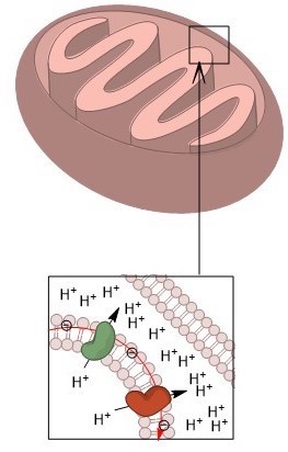
https://upload.wikimedia.org/wikipedia/commons/0/0e/Mitochondria_Intermembrane_pH.jpg
Description English:
A zoomed in snapshot of the proton gradient created across the mitochondrial
inner membrane.
Date 9 June 2019
Source Own work
Author Kayladanesh
https://commons.wikimedia.org/wiki/File:Mitochondria_Intermembrane_pH.jpg
Cells
https://commons.wikimedia.org/wiki/File:Organelles_of_the_Secretory_Pathway.png
https://upload.wikimedia.org/wikipedia/commons/thumb/b/b5/Organelles_of_the_Secretory_Pathway.png/551px-Organelles_of_the_Secretory_Pathway.png
Description English:
This image shows the organelles that make up the pathway of cellular secretion.
Date 31 January 2013, 15:26:33
Source http://open.umich.edu/education/med/resources/second-look-series/materials - Cell Biology Slide 11; Histology of Glands Slide 4
Author Artwork by Holly Fischer
Tissues
https://commons.wikimedia.org/wiki/File:Simple_columnar_epithelium.jpg
https://upload.wikimedia.org/wikipedia/commons/d/dd/Simple_columnar_epithelium.jpg
Description Simple_columnar_epithelium
Date 30 January 2008
Source [1] and Image:Illu epithelium.jpg
Author Made, for the sake of free
knowledge to all mankind, by Mikael Häggström (User:Mikael Häggström)
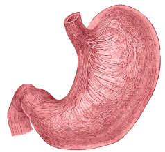
https://commons.wikimedia.org/wiki/File:Histology_of_Stomach.svg
https://upload.wikimedia.org/wikipedia/commons/thumb/c/c9/Histology_of_Stomach.svg/512px-Histology_of_Stomach.svg.png
Description English:
The stomach wall and layers of the digestive tract.
Date 29 March 2020
Source Own work based on: 2415
Histology of StomachN esp.png by OpenStax College
Author Collectivedogs
Organs
https://upload.wikimedia.org/wikipedia/commons/thumb/a/a9/3D_Medical_Animation_Muscular_Layers_of_stomach.jpg/800px-3D_Medical_Animation_Muscular_Layers_of_stomach.jpg
https://commons.wikimedia.org/wiki/File:3D_Medical_Animation_Muscular_Layers_of_stomach.jpg
Description English:
3D Medical Animation still shot of Muscular layers of stomach
Date 15 July 2020
Source https://www.scientificanimations.com/wiki-images/
Author https://www.scientificanimations.com
https://upload.wikimedia.org/wikipedia/commons/thumb/8/8b/Blausen_0604_LargeIntestine2_eu.svg/750px-Blausen_0604_LargeIntestine2_eu.svg.png
https://commons.wikimedia.org/wiki/File:Blausen_0604_LargeIntestine2.png
Description English: The Large Intestine. See a related animation of this medical topic.
Date 8 October 2013, 10:53:10
Source Own
work
Author BruceBlaus.
When using this image in external sources it can be cited as:
Blausen.com
staff (2014). "Medical
gallery of Blausen Medical 2014". wikiJournal of Medicine 1 (2). DOI:10.15347/wjm/2014.010. ISSN 2002-4436.
Permission: This
work is free and may be used by anyone for any purpose. If you wish
to use this content, you do not need
to request permission as long as you follow any licensing requirements
mentioned on this page.
wikimedia
Foundation has received an e-mail confirming that the copyright holder has
approved publication under the terms mentioned on this page. This
correspondence has been reviewed by
an OTRS member and stored in our permission archive. The correspondence is available to trusted
volunteers as ticket #2013061010006654.
If you have
questions about the archived correspondence, please use the OTRS noticeboard.
Ticket link: https://ticket.wikimedia.org/otrs/index.pl?Action=AgentTicketZoom&TicketNumber=2013061010006654
Systems
https://upload.wikimedia.org/wikipedia/commons/1/1d/Stomach.png
Description English:
Organ available for use in the Häggström diagrams
Date 19 September 2010
Source File:Gray1050-stomach.png
Author Mikael Häggström
https://commons.wikimedia.org/wiki/File:Blausen_0316_DigestiveSystem.png
https://upload.wikimedia.org/wikipedia/commons/thumb/1/14/Blausen_0316_DigestiveSystem.png/600px-Blausen_0316_DigestiveSystem.png
Description English: Digestive System. See a full animation of this medical
topic.
Date 5 September
2013, 16:38:38
Source Own work
Author BruceBlaus.
When using this image in external sources it can be cited as:
Blausen.com
staff (2014). "Medical
gallery of Blausen Medical 2014".
wikiJournal of Medicine 1 (2). DOI:10.15347/wjm/2014.010.
ISSN 2002-4436.
Organism
https://commons.wikimedia.org/wiki/File:Female_shadow_anatomy_without_labels.png
https://upload.wikimedia.org/wikipedia/commons/thumb/7/7d/Female_shadow_anatomy_without_labels.png/197px-Female_shadow_anatomy_without_labels.png
Description English: Template for use in Inkscape to make diagrams. Further information at project main
page: Human body
diagrams. To discuss
image, please see Talk:Human body diagrams
Date 22 April
2013
Source All
used images are in public domain.
Author Mikael Häggström
ISSN 2002-4436.
![[1]](http://training.seer.cancer.gov/module_anatomy/images/illu_epithelium.jpg)