Fig. 2.21 Cell life cycle
leading to cell reproduction (a) Life cycle (b) Reproduction by mitosis.
(Sources
of images below. Used with permission.)
Animation:
https://www.youtube.com/watch?v=AhgRhXl7w_g
https://gfycat.com/acrobaticmeaslyamericanblackvulture-chromosomes-cytokinesis-metaphase
Video:
https://en.wikipedia.org/wiki/Mitosis#/media/File:Mitosis_Mesenchymal_Stem_Cells.gif
Copyright
2020: Augustine G. DiGiovanna, Ph.D., Salisbury University, Maryland
The materials on this site are licensed under CC BY-NC-SA 4.0 .

https://www.biologyofhumanaging.com/Figures/CC-BY-NS-SA%20image.jpg
Attribution-NonCommercial-ShareAlike
This license requires that reusers
give credit to the creator. It allows reusers to
distribute, remix, adapt, and build upon the material in any medium or format,
for noncommercial purposes only. If others modify or adapt the material, they
must license the modified material under identical terms.
Previous print editions of the text Human Aging: Biological Perspectives are ©
Copyright 2000, 1994 by The McGraw-Hill Companies, Inc. and 2020 by Augustine
DiGiovanna.
View License Deed | View Legal Code
Sources of image below. Used with permission.

https://upload.wikimedia.org/wikipedia/commons/thumb/e/e0/Cell_Cycle_2-2.svg/587px-Cell_Cycle_2-2.svg.png
https://en.wikipedia.org/wiki/File:Cell_Cycle_2-2.svg
Description English: By Richard Wheeler (Zephyris)
2006. Schematic representation of the cell cycle. Cytokinesis forms rapidly in the process of the
cell cycle.
Date 25
January 2011, 01:43 (UTC)
·
Source Cell_Cycle_2.svg
·
derivative work: Histidine (talk)
This is a retouched
picture, which
means that it has been digitally altered from its original version.
Modifications: changed to better represent duration of cell cycle phases.
The original can be viewed here: Cell
Cycle 2.svg:  . Modifications made by Histidine.
Original figure:
Description English: The four phases of the cell cycle. G1 - the intitial growth phase. S
- the phase in which DNA is synthesised. G2 - the second growth phase in preparation for cell
division. M - mitosis; where the cell divides to produce two daughter cells
that continue the cell cycle.
Date 14
August 2013, 13:25:17
Source Own work
Author Simon Caulton
https://upload.wikimedia.org/wikipedia/commons/thumb/c/c9/Mitosis_Stages.svg/800px-Mitosis_Stages.svg.png
https://commons.wikimedia.org/wiki/File:Mitosis_Stages.svg
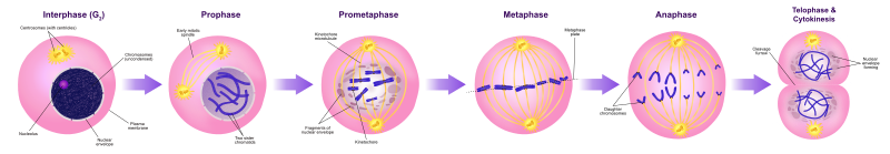
Description English: A diagram of mitosis stages
Interphase (G₂): In this substage, the cell prepares for nuclear
division and a protein that makes microtubles for
cell division is synthesized.
Prophase: The longest stage of mitosis. In this stage the chromosomes
become visible and the centrioles separate and move to opposite poles of the cell.
Prometaphase: The nuclear envelope disintegrates and microtubules can
attach to kinetochores. Chromosomes congress toward the metaphase plate of the
cell.
Metaphase: In this stage the chromosomes line up across the center of
the cell and become connected to the spindle fiber at their centromere.
Anaphase: In this stage the sister chromatids separate into individual
chromosomes and are pulled apart.
Telophase & cytokinesis: Chromosomes decondense and are surrounded
by a newly formed nuclear envelope. Cytokinesis typically coincides with and
telophase.
Date 26
June 2016
Source Own work; Used information from: Campbell Biology (10th
Edition) by: Jane B. Reece & Steven A. Wasserman.
and Nature.com.
Author Ali Zifan
Animations
https://www.youtube.com/watch?v=AhgRhXl7w_g
https://gfycat.com/acrobaticmeaslyamericanblackvulture-chromosomes-cytokinesis-metaphase
Video
https://en.wikipedia.org/wiki/Mitosis#/media/File:Mitosis_Mesenchymal_Stem_Cells.gif