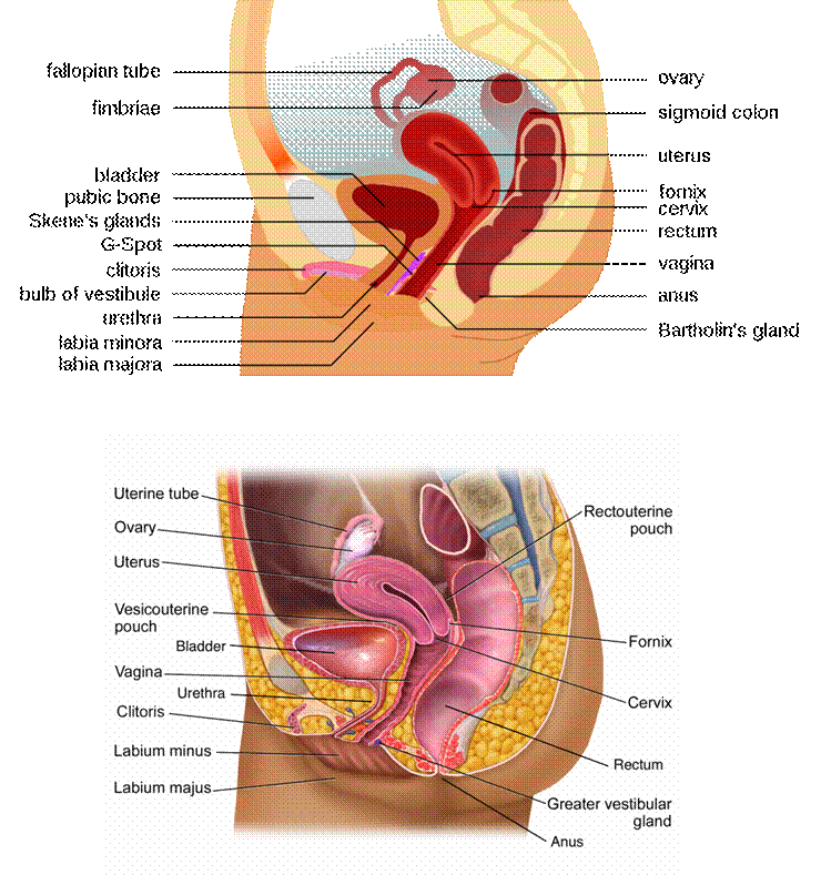
Fig. 13.7 Structure of the female reproductive system: Mid-sagittal
section of female pelvis - two versions. (Sources of images and videos below. Used
with permission.)
Videos
Female system
“Reproductive System (Female)”
https://blausen.com/en/video/reproductive-system-female/
“The Function of Ovaries”
https://blausen.com/en/video/the-function-of-ovaries/
“The Ovaries and Ovulation”
https://blausen.com/en/video/the-ovaries-and-ovulation/
“Ovulation”
https://blausen.com/en/video/ovulation/
“Oocyte (Egg)”
https://blausen.com/en/video/oocyte-egg/
©
Copyright 2020: Augustine G. DiGiovanna, Ph.D.,
Salisbury University, Maryland
The materials on this site are licensed under CC BY-NC-SA
4.0
![]()
Attribution-NonCommercial-ShareAlike
This license requires that reusers
give credit to the creator. It allows reusers
to distribute, remix, adapt, and build upon the material in any medium
or format, for noncommercial purposes only. If others modify or adapt
the material, they must license the modified material under identical
terms.
Previous print editions of the text Human Aging: Biological Perspectives
are © Copyright 2000, 1994 by The McGraw-Hill Companies, Inc. and 2020
by Augustine DiGiovanna.
View License Deed |
View Legal Code
Sources of images and videos. Used with permission.

https://commons.wikimedia.org/wiki/File:Female_anatomy_with_g-spot-en.svg
Description English:
lateral anatomy view of the female reproductive system with English
terms (with the G-Spot)
Date 9 January 2010
Source Own work
Author Tsaitgaist
Other versions
SVG development The
source code of this SVG is valid. This
diagram was created with Inkscape, or with something else. This SVG diagram uses embedded
text that can be easily
translated using a text editor.
This is a retouched picture, which means that it has
been digitally altered from its original version. Modifications: vectorization.
The original can be viewed here: Female anatomy.svg: File:Female anatomy.svg. Modifications made by Tsaitgaist.
Licensing
I, the copyright holder of this work, hereby publish it under the following
licenses:
This file is licensed under the Creative Commons Attribution-Share Alike 3.0 Unported license.
You are free:
·
to share – to copy, distribute and transmit the work
·
to remix – to adapt the work
Under the following conditions:
·
attribution – You must give appropriate credit, provide a link to the license, and
indicate if changes were made. You may do so in any reasonable manner, but not
in any way that suggests the licensor endorses you or your use.
share alike – If you remix, transform,
or build upon the material, you must distribute your contributions under the same or compatible license as the original.
Permission is granted to copy, distribute and/or modify this
document under the terms of the GNU Free Documentation License, Version 1.2 or any later version published by
the Free Software Foundation; with no Invariant Sections, no Front-Cover Texts, and no Back-Cover
Texts. A copy of the license is included in the section entitled GNU Free Documentation License.
You may select the license of your choice.

https://commons.wikimedia.org/wiki/File:Blausen_0400_FemaleReproSystem_02b.png
Description English: Female Reproductive System (Sectional
view). See a full animation of this medical topic. Additional callouts added
and caption removed.
Русский: Женская
репродуктивная
система (в
разрезе, вид
сбоку)
Date 14 February 2017
Source This file
was derived from: Blausen 0400 FemaleReproSystem 02.png
Author BruceBlaus.
When using this image in external sources it can be cited as:
·
Blausen.com staff (2014). "Medical gallery of Blausen Medical 2014". WikiJournal of Medicine 1 (2). DOI:10.15347/wjm/2014.010. ISSN 2002-4436.
Modified by User:ArnoldReinhold who
released mods under CC0
Permission
(Reusing this file) This work is free and may be used by anyone
for any purpose. If you wish to use this content, you do not need to request permission as long
as you follow any licensing requirements mentioned on this page.
The Wikimedia
Foundation has received an e-mail confirming that the copyright holder has
approved publication under the terms mentioned on this page. This
correspondence has been reviewed by
an Volunteer Response Team (VRT) member and stored in our permission archive. The correspondence is available to trusted
volunteers ticket #2013061010006654.
If you have questions about the archived correspondence, please use the OTRS noticeboard. Ticket link: https://ticket.wikimedia.org/otrs/index.pl?Action=AgentTicketZoom&TicketNumber=2013061010006654
See source file File:Blausen 0400 FemaleReproSystem 02.png
Other versions
Licensing
The copyright holder of this
file allows anyone to use it for any purpose, provided that the
copyright holder is properly attributed. Redistribution, derivative work,
commercial use, and all other use is permitted.
Videos
Female system
“Reproductive System (Female)”
https://blausen.com/en/video/reproductive-system-female/
“The Function of Ovaries”
https://blausen.com/en/video/the-function-of-ovaries/
“The Ovaries and Ovulation”
https://blausen.com/en/video/the-ovaries-and-ovulation/
“Ovulation”
https://blausen.com/en/video/ovulation/
“Oocyte (Egg)”
https://blausen.com/en/video/oocyte-egg/