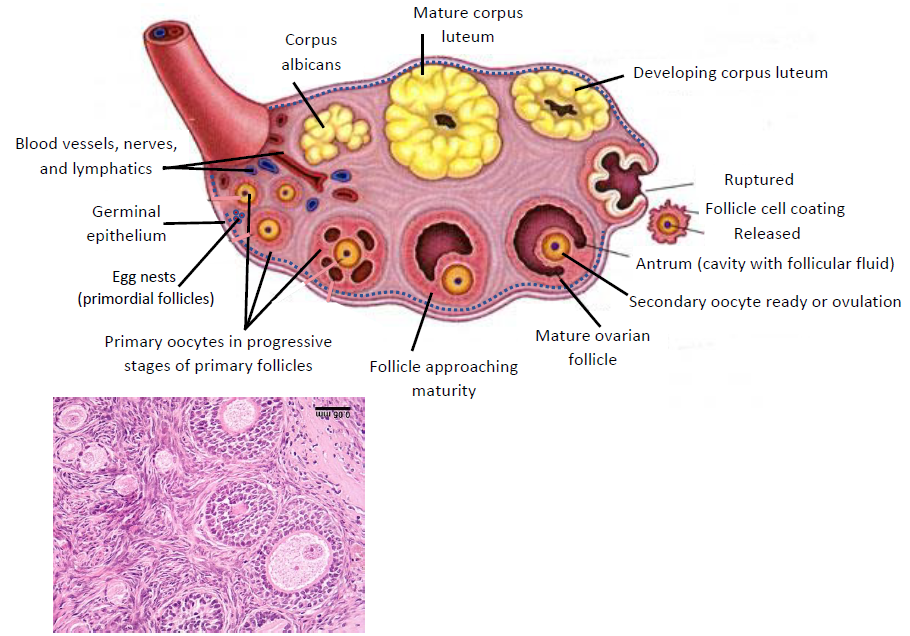
Fig. 13.10 Ovarian structure, follicle
development, and ovulation. (Sources of images and videos below. Used with
permission.)
Videos
“Reproductive System (Female)”
https://blausen.com/en/video/reproductive-system-female/
“The Function of Ovaries”
https://blausen.com/en/video/the-function-of-ovaries/
“The Ovaries and Ovulation”
https://blausen.com/en/video/the-ovaries-and-ovulation/
“Ovulation”
https://blausen.com/en/video/ovulation/
“Oocyte (Egg)”
https://blausen.com/en/video/oocyte-egg/
©
Copyright 2020: Augustine G. DiGiovanna, Ph.D.,
Salisbury University, Maryland
The materials on this site are licensed under CC BY-NC-SA
4.0
![]()
Attribution-NonCommercial-ShareAlike
This license requires that reusers
give credit to the creator. It allows reusers
to distribute, remix, adapt, and build upon the material in any medium
or format, for noncommercial purposes only. If others modify or adapt
the material, they must license the modified material under identical
terms.
Previous print editions of the text Human Aging: Biological Perspectives
are © Copyright 2000, 1994 by The McGraw-Hill Companies, Inc. and 2020
by Augustine DiGiovanna.
View License Deed |
View Legal Code
Sources of images and videos. Used with permission.

https://commons.wikimedia.org/wiki/File:Anatomy_of_the_ovaries.jpg
Description English: Anatomy
of the internal structures of the ovary.
Date 21 July 2018
Author Kimanh Nguyen
Licensing
This file is licensed under
the Creative Commons Attribution-Share Alike 3.0 Unported license.
You are free:
·
to share – to copy, distribute and transmit the work
·
to remix – to adapt the work
Under the following conditions:
·
attribution – You must give appropriate credit, provide a link to the license, and
indicate if changes were made. You may do so in any reasonable manner, but not
in any way that suggests the licensor endorses you or your use.
share alike – If you remix, transform, or
build upon the material, you must distribute your contributions under the same or compatible license as the original.
Modified: A.G.
DiGIovanna
Changed labels. Added labels
and lines.

https://en.wikipedia.org/wiki/File:Ovarian_cortex_in_a_rhesus_monkey.jpg
Description
Micrograph
of the ovarian cortex showing several round follicles embedded in a matrix of
stromal cells. A mature follicle is at the upper left. The ovary was cut at a
thickness of 10 microns (0.01 mm) and stained with the dyes
hematoxylin and eosin.
Source
Photo
of a histologic section taken with a microscope
Date
2020-05-06
Author
Tulemo
Permission
(Reusing
this file)
See
below.
Licensing
I, the copyright holder of this work, hereby publish it under the following licenses:
Permission is granted to copy, distribute and/or modify this document
under the terms of the GNU Free Documentation License, Version
1.2 or any later version published by the Free Software Foundation; with no
Invariant Sections, no Front-Cover Texts, and no Back-Cover Texts.
This work is licensed under the
Creative Commons Attribution-ShareAlike 4.0 License.
You may select the license of your choice.
Videos
“Reproductive System (Female)”
https://blausen.com/en/video/reproductive-system-female/
“The Function of Ovaries”
https://blausen.com/en/video/the-function-of-ovaries/
“The Ovaries and Ovulation”
https://blausen.com/en/video/the-ovaries-and-ovulation/
“Ovulation”
https://blausen.com/en/video/ovulation/
“Oocyte (Egg)”
https://blausen.com/en/video/oocyte-egg/