Fig. 12.2 Kidney structure. (Sources of images and videos below. Used
with permission.)
Videos
“Overview of the Urinary System”
https://blausen.com/en/video/overview-of-the-urinary-system/
©
Copyright 2020: Augustine G. DiGiovanna, Ph.D.,
Salisbury University, Maryland
The materials on this site are licensed under CC BY-NC-SA
4.0
![]()
Attribution-NonCommercial-ShareAlike
This license requires that reusers
give credit to the creator. It allows reusers
to distribute, remix, adapt, and build upon the material in any medium
or format, for noncommercial purposes only. If others modify or adapt
the material, they must license the modified material under identical
terms.
Previous print editions of the text Human Aging: Biological Perspectives
are © Copyright 2000, 1994 by The McGraw-Hill Companies, Inc. and 2020
by Augustine DiGiovanna.
View License Deed |
View Legal Code
Sources of images and videos below. Used with permission.
https://commons.wikimedia.org/wiki/File:Blausen_0592_KidneyAnatomy_01.png
Description English: Kidney Anatomy. See a related animation of this medical topic.
Date 11 February 2014, 12:55:15
Source Own work
Author BruceBlaus.
When using this image in external sources it can be cited as:
·
Blausen.com staff
(2014). "Medical
gallery of Blausen Medical 2014".
WikiJournal of Medicine 1 (2). DOI:10.15347/wjm/2014.010. ISSN 2002-4436.
Permission
(Reusing this file) This work
is free and may be used by anyone
for any purpose. If you wish to use this content, you do not need to request permission as long as you
follow any licensing requirements mentioned on this page.
The Wikimedia Foundation has received an e-mail
confirming that the copyright holder has approved publication under the terms
mentioned on this page. This correspondence has been reviewed by an OTRS member and stored in our permission archive. The correspondence is
available to trusted volunteers as ticket #2013061010006654.
If you have questions about
the archived correspondence, please use the OTRS noticeboard. Ticket link: https://ticket.wikimedia.org/otrs/index.pl?Action=AgentTicketZoom&TicketNumber=2013061010006654
This
work is free and may be used by anyone
for any purpose. If you wish to use this content, you do not need to request permission as long as you
follow any licensing requirements mentioned on this page.
The Wikimedia Foundation has received an e-mail
confirming that the copyright holder has approved publication under the terms
mentioned on this page. This correspondence has been reviewed by an OTRS member and stored in our permission archive. The correspondence is
available to trusted volunteers as ticket #2013061010006654.
If you have questions about
the archived correspondence, please use the OTRS noticeboard. Ticket link: https://ticket.wikimedia.org/otrs/index.pl?Action=AgentTicketZoom&TicketNumber=2013061010006654
This biology image could be re-created using
vector graphics as an SVG file. This has several
advantages; see Commons:Media for cleanup for more
information. If an SVG form of this image is
available, please upload it and afterwards replace this template with {{vector version available|new image name}}.
It is recommended to name the SVG file "Blausen
0592 KidneyAnatomy 01.svg" – then the template Vector version available (or Vva) does not need the new image name parameter.
Licensing
I, the copyright holder of
this work, hereby publish it under the following license:
This file is licensed under
the Creative Commons Attribution 3.0 Unported license.
You
are free:
·
to share – to copy, distribute and transmit the work
·
to remix – to adapt the work
Under the following conditions:
attribution – You must give appropriate
credit, provide a link to the license, and indicate if changes were made. You
may do so in any reasonable manner, but not in any way that suggests the
licensor endorses you or your use.
Modified: A.G. DiGiovanna
Added labels and lines.
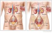
https://www.ncbi.nlm.nih.gov/books/NBK65775/bin/CDR0000765031.jpg
“Bladder
and Other Urothelial Cancers Screening (PDQ®)”
Patient Version
PDQ Screening and
Prevention Editorial Board.
Published online: August 19, 2020.
https://www.ncbi.nlm.nih.gov/books/NBK65775/
https://www.ncbi.nlm.nih.gov/books/NBK65775/bin/CDR0000765031.jpg
Modified: A.G. DiGiovanna
Added labels and lines. Blocked some
portions.
Videos
“Overview of the Urinary System”
https://blausen.com/en/video/overview-of-the-urinary-system/