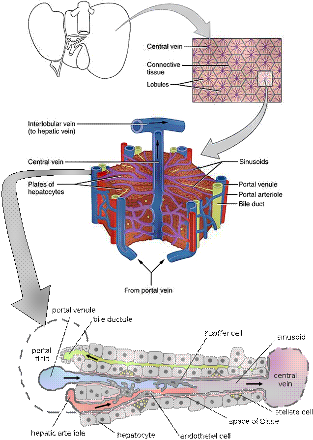
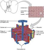
Fig. 10.6 Structure of liver lobules.
(Sources of images and videos below. Used with permission.)
Videos
“Liver Function”
https://blausen.com/en/video/liver-function/
“Liver Cirrhosis”
https://blausen.com/en/video/liver-cirrhosis/
“Hepatitis”
https://blausen.com/en/video/hepatitis/
“Gallstones”
https://blausen.com/en/video/gallstones/
“Carbohydrate Metabolism in
the Liver”
https://blausen.com/en/video/carbohydrate-metabolism-in-the-liver/
©
Copyright 2020: Augustine G. DiGiovanna, Ph.D.,
Salisbury University, Maryland
The materials on this site are licensed under CC BY-NC-SA
4.0
![]()
Attribution-NonCommercial-ShareAlike
This license requires that reusers
give credit to the creator. It allows reusers
to distribute, remix, adapt, and build upon the material in any medium
or format, for noncommercial purposes only. If others modify or adapt
the material, they must license the modified material under identical
terms.
Previous print editions of the text Human Aging: Biological Perspectives
are © Copyright 2000, 1994 by The McGraw-Hill Companies, Inc. and 2020
by Augustine DiGiovanna.
View License Deed |
View Legal Code
Sources of images and videos. Used with permission.
https://upload.wikimedia.org/wikipedia/commons/b/b7/2423_Microscopic_Anatomy_of_Liver.jpg
Description Illustration from
Anatomy & Physiology, Connexions Web site. http://cnx.org/content/col11496/1.6/, Jun 19, 2013.
Source Anatomy &
Physiology, Connexions Web site. http://cnx.org/content/col11496/1.6/, Jun 19, 2013.
Author OpenStax College
Other versions
Licensing
This work is ineligible for copyright and therefore in
the public domain because it consists entirely of information that is common property and
contains no original authorship.
Modified: A.G. DiGiovanna
Added arrow.

https://upload.wikimedia.org/wikipedia/commons/a/a0/Cellular_architecture_of_the_liver.jpg
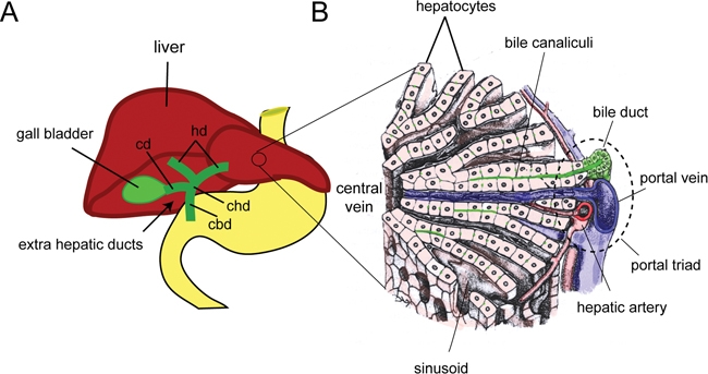
Description English:
(A) The schematic shows an adult liver (red), with the gall bladder
and extra hepatic ducts (green), in relation to the stomach and intestine
(yellow). The extra hepatic duct system consists of the hepatic ducts (hd), which drain bile from the liver into the common
hepatic duct (chd) to the gall bladder via the cystic
duct (cd) and into the duodenum through the common bile duct (cbd). (B) A schematic of the cellular architecture of the
liver showing the hepatocytes (pink) arranged in hepatic plates separated by
sinusoid spaces radiating around a central vein. Bile canaliculi on the surface
of adjoining hepatocytes drain bile into the bile ducts (green), which run
parallel to portal veins (blue) and hepatic arteries (red) to form the “portal
triad”. (Panel B is adapted with permission from Bloom and Fawcett: A Text Book
of Histology 10th Edition).
Date Published October 31,
2008.
Source [1] Direct
StemBook Figure 3 Cellular architecture of the
liver.
·
Zorn, A.M., Liver development (October 31, 2008), StemBook,
ed. The Stem Cell Research Community, StemBook, doi/10.3824/stembook.1.25.1, http://www.stembook.org.
Author Zorn, A.M., Liver
development (October 31, 2008), StemBook, ed. The
Stem Cell Research Community, StemBook, doi/10.3824/stembook.1.25.1, http://www.stembook.org.
Permission
(Reusing this file)
This file is
licensed under the Creative Commons Attribution 3.0 Unported license.
You are free:
·
to share – to copy, distribute and transmit the work
·
to remix – to adapt the work
Under the following
conditions:
attribution – You must give appropriate credit, provide a
link to the license, and indicate if changes were made. You may do so in any
reasonable manner, but not in any way that suggests the licensor endorses you
or your use.
https://commons.wikimedia.org/wiki/File:Hepatic_structure2.svg
Description English: Illustration of part of a mammalian liver
lobule
Source Based on the
research article "Intravital Observation of Plasmodium berghei Sporozoite Infection of the Liver", PLoS Biology, doi:10.1371/journal.pbio.0030192.g011
Author Originally by Frevert U, Engelmann S, Zougbédé
S, Stange J, Ng B, et al. Converted to SVG by
Viacheslav Vtyurin who was hired to do so by User:Eug.
Other versions Arabic
SVG development The source code of this SVG is valid. This vector
image was created with Adobe Illustrator.
{| cellspacing="8"
cellpadding="0" style="clear:both;
width:100%; margin:0.5em auto; border:2px solid #8888aa; background-color:#f7f8ff;"
lang="en" dir="ltr"
|align="center"|
|width="100%"| This SVG file contains embedded text that can be
translated into your language, using any capable SVG editor, text editor or
the SVG Translate tool. For more information see: About
translating SVG files.
|}
Licensing
This file is licensed under the Creative Commons Attribution 2.5 Generic license.
You are free:
·
to share – to copy, distribute and transmit the work
·
to remix – to adapt the work
Under the following
conditions:
attribution – You must give appropriate credit, provide a
link to the license, and indicate if changes were made. You may do so in any reasonable
manner, but not in any way that suggests the licensor endorses you or your use.
This file was
published in a Public Library of
Science journal. Their website states that the content of all PLOS journals is published under the Creative
Commons Attribution 4.0 license (or its previous version depending on the
publication date), unless indicated otherwise.
Videos
“Liver Function”
https://blausen.com/en/video/liver-function/
“Liver Cirrhosis”
https://blausen.com/en/video/liver-cirrhosis/
“Hepatitis”
https://blausen.com/en/video/hepatitis/
“Gallstones”
https://blausen.com/en/video/gallstones/
“Carbohydrate Metabolism in
the Liver”
https://blausen.com/en/video/carbohydrate-metabolism-in-the-liver/