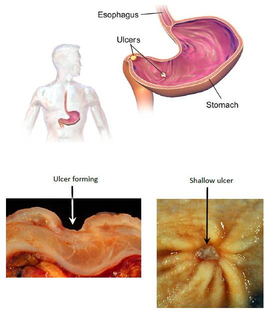
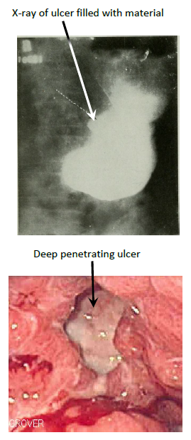
Fig. 10.3 Peptic ulcers in the stomach
and duodenum. (Sources of images and videos below. Used with permission.)
Videos
“Gastric
Ulcers”
https://blausen.com/en/video/gastric-ulcers/
©
Copyright 2020: Augustine G. DiGiovanna, Ph.D.,
Salisbury University, Maryland
The materials on this site are licensed under CC BY-NC-SA
4.0
![]()
Attribution-NonCommercial-ShareAlike
This license requires that reusers
give credit to the creator. It allows reusers
to distribute, remix, adapt, and build upon the material in any medium
or format, for noncommercial purposes only. If others modify or adapt
the material, they must license the modified material under identical
terms.
Previous print editions of the text Human Aging: Biological Perspectives
are © Copyright 2000, 1994 by The McGraw-Hill Companies, Inc. and 2020
by Augustine DiGiovanna.
View License Deed |
View Legal Code
Sources of images
and videos. Used with permission.
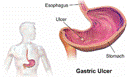
https://upload.wikimedia.org/wikipedia/commons/8/8a/Gastric_Ulcer.png
Description English:
Gastric Ulcer. See a full animation of this medical topic.
Date 10 November 2015
Source Own
work
Author BruceBlaus
Permission
(Reusing this file) This work is free and may be used by anyone for any purpose. If
you wish to use this content, you do not need to
request permission as long as you follow any licensing requirements mentioned
on this page.
wikimedia
Foundation has received an e-mail confirming that the copyright holder has
approved publication under the terms mentioned on this page. This
correspondence has been reviewed by
an OTRS member and stored in our permission
archive. The correspondence is available to trusted
volunteers ticket
#2013061010006654.
If you have questions about the
archived correspondence, please use the OTRS noticeboard. Ticket link: https://ticket.wikimedia.org/otrs/index.pl?Action=AgentTicketZoom&TicketNumber=2013061010006654
Licensing
I, the copyright
holder of this work, hereby publish it under the following license:
This file is licensed under the Creative commons Attribution-Share Alike 4.0 International license.
You are free:
·
to share – to copy, distribute and transmit the work
·
to remix – to adapt the work
Under the following
conditions:
·
attribution – You must give appropriate credit, provide a link to the license, and
indicate if changes were made. You may do so in any reasonable manner, but not
in any way that suggests the licensor endorses you or your use.
share alike – If you remix, transform, or build upon the
material, you must distribute your contributions under the same or compatible license as the original.
Modified: A.G.DiGiovanna
Added componenets, labels, lines, and blocked
label
_(14755473894).jpg)
Description English:
Identifier:
americanjournroen08ameruoft (find
matches)
Title: The
American journal of roentgenology, radium therapy and nuclear medicine
Year: 1906 (1900s)
Authors: American
Radium Society American
Roentgen Ray Society
Subjects: Radiotherapy X-rays
Publisher: Springfield,
Ill. C.C. Thomas
Contributing Library: Gerstein
- University of Toronto
Digitizing Sponsor: University
of Toronto
View Book Page: Book
Viewer
About This Book: Catalog Entry
View All Images: All
Images From Book
Click here to view book
online to see this illustration in context in a browseable online version of this book.
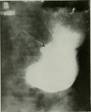
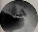
Text Appearing Before Image:
Fig. 13. Case 154564. Roentgenogram. Niche-type ofulcer,
benign. Arrow points to niche. Compare withFigure 14.
Fig. 15. Case 20799. Roentgenogram. Niche-type ofulcer.
Niche extraordinarily large. Operation. Can-cer
demonstrated microscopically.
Text Appearing After Image:
Fig. 14. Case 116521. Roentgenogram. Niche-type ofulcer,
malignant. Arrow points to niche. BIBLIOGRAPHY 1. .\ii\Mi,
J. G., AND NiCHOLLS, A. G. Principles of pathology.
Philadelphia, 1909, ii, p. 417. 2. AscHOFF, L. Ueber die mechanischen Momente in der Pathogenese des rundcn Magengeschwiirsund iibcr seine Beziehungen zum Krebs. Deutsche, vied. IVchuschr.,
Berl., 1912, xxxviii,494-496. 3. AscHOFF,
L. Pathologische Anatomic. Jena, 1919, ii, 824; 839.
4. AssMAN, H. Die Roentgendiagnostik
der inneren Erkrankungen.
Leipzig, 1921, 385 ; 419; 693. 5. Bland-Sutton,
J. Ulcers new and old; jejunal for duodenal ulcers. Lancet, 1916, i, 387-391. 6. Carman, R. D. A new roentgen-ray sign of ul-
cerating gastric cancer. /. Am. M. Assn. 1921,Ixxvii, 990-992. 7. Cheney, W. F. Gastric cancer as a
sequel to gastric ulcer. /. Am. M. Assn., 1915, Ixv,
1227-1231. 8. Clairmont P., AND HaudeIk, M. Die Bedeutung der Magenradiologie fiir die Chirurgie. Jena, 1911,
P- 53- 9. Deaver, J. B. Gastric ulcer. Am. J.
Note About Images
Please note that these images are extracted from scanned page images that
may have been digitally enhanced for readability - coloration and appearance of
these illustrations may not perfectly resemble the original work.
Date 1906
Source https://www.flickr.com/photos/internetarchivebookimages/14755473894/
·
Source book page: https://archive.org/stream/americanjournroen08ameruoft/americanjournroen08ameruoft#page/n721/mode/1up
Author Internet Archive Book Images
Permission
(Reusing this file) At the time of upload, the image license
was automatically confirmed using the Flickr API. For more information see Flickr API detail.
·
Flickr tags bookid:americanjournroen08ameruoft
This image was taken
from Flickr's The commons. The uploading
organization may have various reasons for determining that no known
copyright restrictions exist, such as:
1.
The copyright is in
the public domain because it has expired;
2.
The copyright was injected
into the public domain for other reasons, such as failure to adhere to required
formalities or conditions;
3.
The institution owns
the copyright but is not interested in exercising control; or
4.
The institution has
legal rights sufficient to authorize others to use the work without
restrictions.
More
information can be found at https://flickr.com/commons/usage/.
Please add
additional copyright tags to this image if more specific information about
copyright status can be determined. See commons:Licensing for more information.
This image was originally posted to
Flickr by Internet Archive Book Images at https://flickr.com/photos/126377022@N07/14755473894. It was reviewed on 16 September 2015 by FlickreviewR and was confirmed to be licensed under the terms of the No known copyright
restrictions.
Modifed: A.G. DiGiovanna
Added labels and
lines.
https://commons.wikimedia.org/wiki/File:Gastric_ulcer_3.jpg
Description English:
Endoscopic image of gastric ulcer, biopsy proven to be en:gastric cancer. Released into public domain on permission of
patient -- Samir धर्म 08:44, 7 June 2006 (UTC)
Date 7 June 2006 (original
upload date)
Source Transferred from en.wikipedia to commons.
Author Samir (The Scope) at English wikipedia
Licensing
Samir (The Scope) at the English wikipedia, the copyright
holder of this work, hereby publishes it under the following license:
Permission is
granted to copy, distribute and/or modify this document under the terms of the GNU Free
Documentation License, Version 1.2 or any
later version published by the Free Software
Foundation; with no Invariant Sections, no Front-Cover
Texts, and no Back-Cover Texts. A copy of the license is included in the
section entitled GNU Free Documentation License.
This file is licensed under the Creative commons Attribution-Share Alike 3.0 Unported license.
Attribution: Samir (The Scope) at the English wikipedia
You are free:
·
to share – to copy, distribute and transmit the work
·
to remix – to adapt the work
Under the following
conditions:
·
attribution – You must give appropriate credit, provide a link to the license, and
indicate if changes were made. You may do so in any reasonable manner, but not
in any way that suggests the licensor endorses you or your use.
share alike – If you remix, transform, or build upon the
material, you must distribute your contributions under the same or compatible license as the original.
This licensing tag was added to this file as part of the GFDL licensing update.
Modifed: A.G. DiGiovanna
Added labels and
lines.
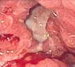
https://commons.wikimedia.org/wiki/File:Benign_gastric_ulcer_3.jpg
Description Benign gastric ulcer
This photo presents the same case
shown in Image:Benign gastric ulcer 1.jpg and Image:Benign gastric ulcer 2 (wet).jpg.
.jpg)
The photo is a of a longitudinal
section of the ulcer and adjacent gastric wall. Note that the normal anatomic
layers are discrete and relatively undisturbed as compared with the homogenation of the wall and caused by some malignant
ulcers.
Photograph by Ed Uthman, MD. Public
domain. Posted 23 Sep 00
Source http://web2.airmail.net/uthman/specimens/index.html
Author Ed Uthman, MD.
Permission
(Reusing this file) PD
Summary
This work has been released into the public domain by its author, Ed
Uthman. This applies worldwide.
In some countries this may not be legally
possible; if so:
Ed Uthman grants anyone the right to use this work for any purpose,
without any conditions, unless such conditions are required by law.
Modifed: A.G. DiGiovanna
Added labels and
lines.
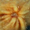
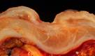
https://commons.wikimedia.org/wiki/File:Benign_gastric_ulcer_1.jpg
Description Čeština: Antrální
žaludeční vřed (histologicky benigní)
English: gastric ulcer
This 1-cm
benign gastric antral ulcer was discovered serendipitously in a gastrectomy
specimen removed for adenocarcinoma of the fundus (not shown in the photo). The
gross appearance is classic for a benign ulcer in that 1) it is relatively
small, 2) the mucosa surrounding the ulcer base does not appear tumefactive, and 3) the radiating rugal
folds extend nearly all the way to the margins of the base. Contrast this
appearance with that of the malignant gastric ulcer included in this case
collection. The criteria for grossly and endoscopically distinguishing benign
ulcers from cancer are not absolute, which is why it is necessary to perform a
biopsy on any non-healing gastric ulcer. Even biopsies are not 100% accurate in
picking up a cancer, so negative pathology reports in such cases may provide
false reassurance.
The photo was taken with a Minolta X-370 with 100mm bellows lens on Kodak
Elite ISO 100 film. The specimen was previously fixed overnight in formalin
after being pinned out in a wax-bottomed tray.
Date Posted 23 Sep 00
Source http://web2.airmail.net/uthman/specimens/index.html
Author Ed Uthman, MD
Permission
(Reusing this file) This work has been
released into the public domain by its author, Ed
Uthman. This applies worldwide.
In some countries this may not be legally
possible; if so:
Ed Uthman grants anyone the right to use this work for any purpose,
without any conditions, unless such conditions are required by law.
Modifed: A.G. DiGiovanna
Added labels and
lines.
Videos
“Gastric
Ulcers”
https://blausen.com/en/video/gastric-ulcers/