Inflammation - Chronic gastritis
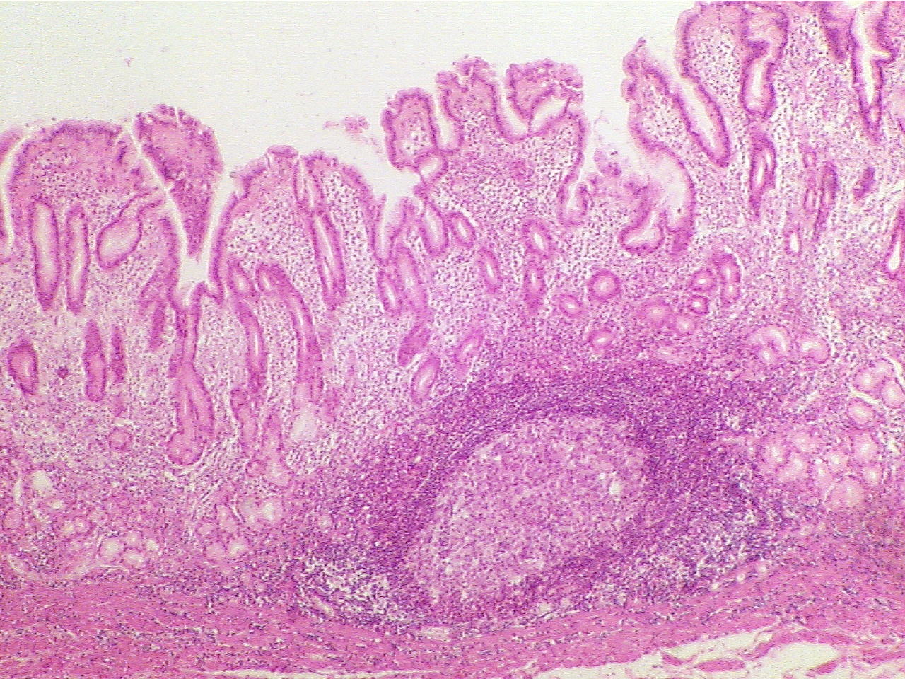
Compare the normal stomach mucosa with the slide showing gatritis. Notice that on the slide of gastritis, the mucosa is thinned and flattened and there is exudate (clear spaces with pink structureless material) present within the submucosa. Notice also that the lymph nodes are greatly enlarged and there are large numbers of white blood cells (stained blue) within the submucosa.
Normal stomach (40X2.8) Gastritis (40X2.8)
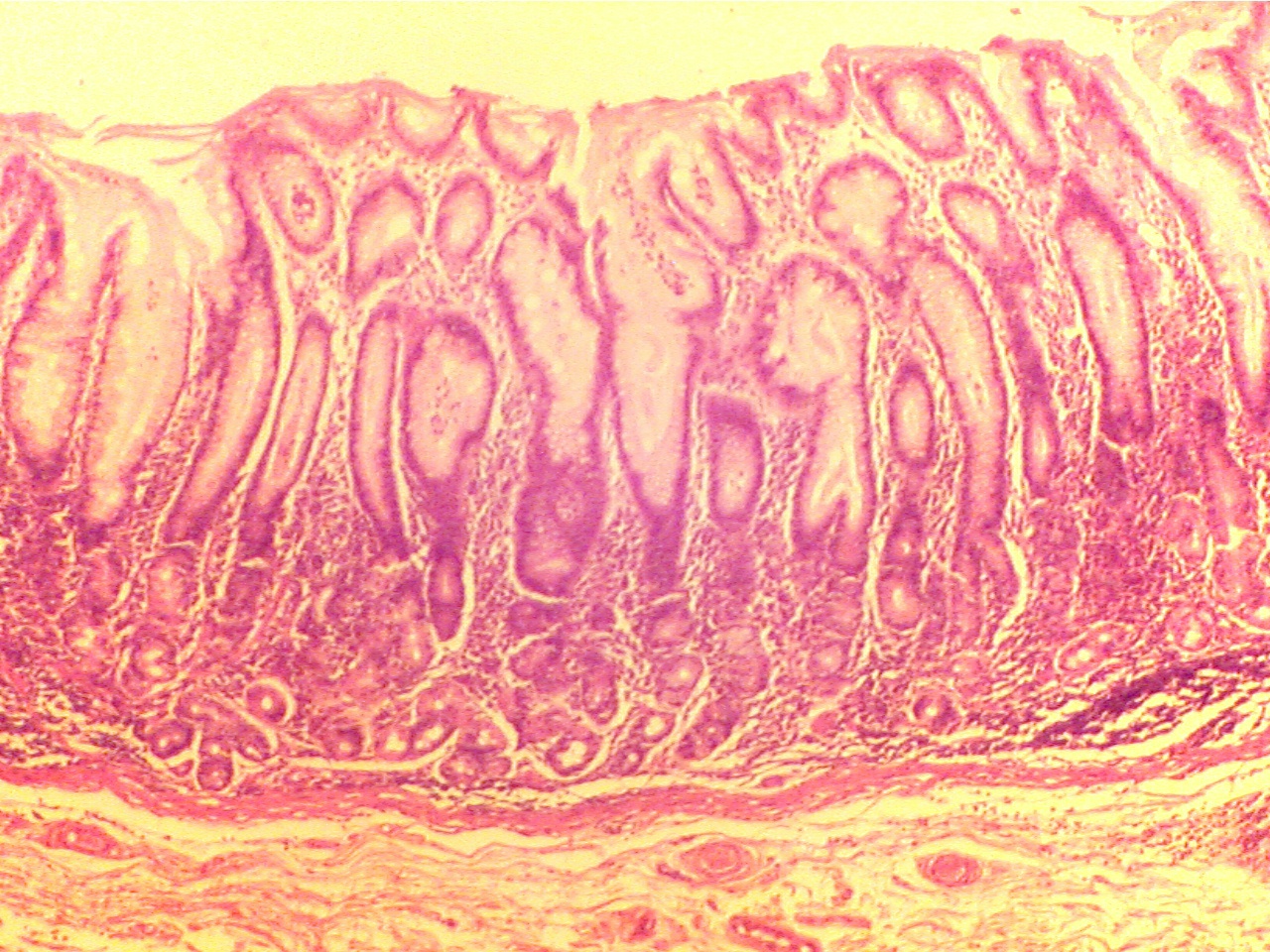
Mucosa is dark red layer on upper surface and Mucosa is dark layer on upper surface and lining
lining pit-like vertical glands. Submucosa is pit-like glands. The glands are widely separated
granular material between glands. by the exudate-filled submucosa. Large lymph node
at base of submucosa is surrounded by WBCs.
Normal stomach (100X2.8) Gastritis (400X2.8)
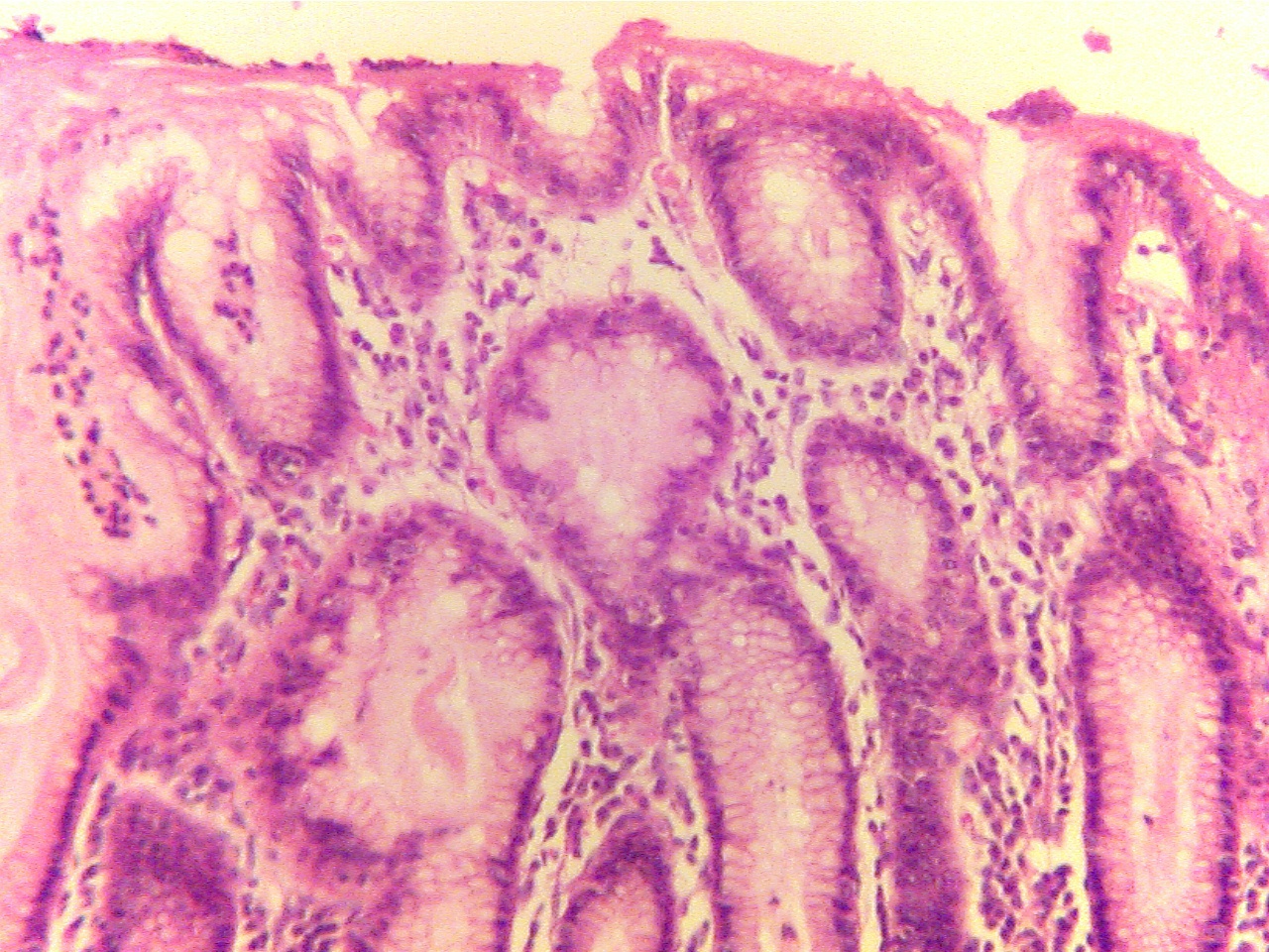
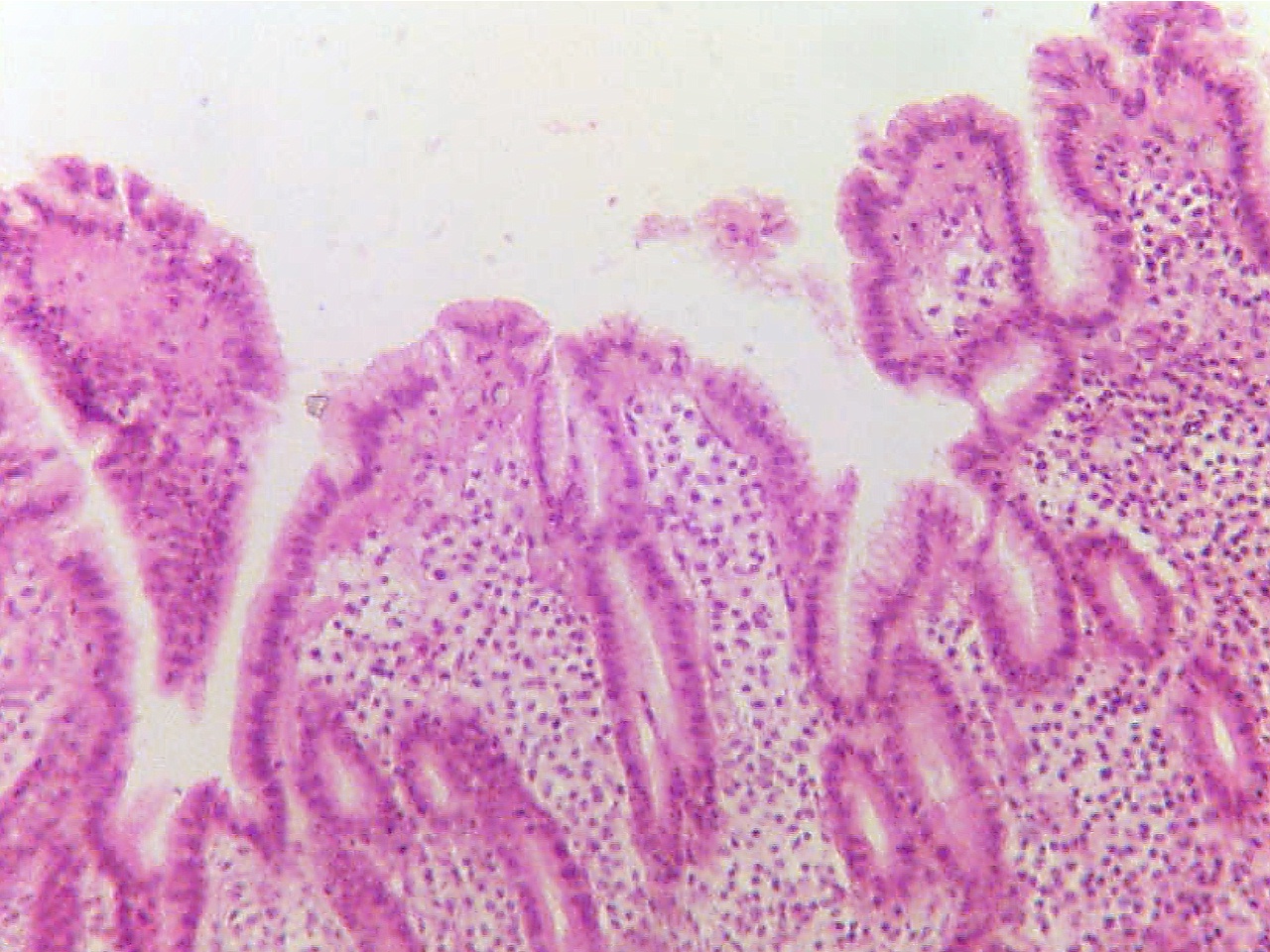
Mucosa is dark layer on upper surface and Mucosa is dark layer on upper surface and lining
lining pit-like vertical glands. Submucosa is pit-like glands. The glands are widely separated
scattered cells in clear gel between glands by the exudate-filled submucosa, which contains.
many small dark inflammatory cells (WBCs).
Return to Slide List